Valgus slipped capital femoral epiphysis
- PMID: 21045997
- PMCID: PMC2958296
Valgus slipped capital femoral epiphysis
Abstract
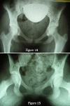
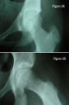
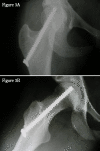
Valgus slips of the epiphysis are rare, making radiological diagnosis difficult. A high degree of clinical suspicion is required to diagnose the condition. The patient was a 13-year, 7-month-old girl who had been suffering from pain in the left thigh for ten days. She had a limp and a positive Trendelenburg sign. Menstrual function had started when she was 12 years and 10 months old. Pain occurred with getting up from a chair. Hip radiographs revealed symmetrical, bilateral caput valgum, which was a potential cause of confusion given the valgus displacement of the proximal femoral epiphysis. Axial view showed an almost imperceptible posterior slip. The patient was diagnosed as having a valgus slipped capital femoral epiphysis (SCFE). Surgical treatment was performed using in-situ fixation with a cannulated, fully threaded percutaneous screw placed through the external cortex of the femoral neck. Non-weight-bearing for six weeks was prescribed. Although a medial approach is usually used for screw insertion using a more medial entry-point, preventing neurovascular risks, in-situ fixation (through a lateral approach) was performed more safely and distally. This was done through the outer cortex of the femoral neck (and centered in the axial view), to achieve fixation of the femoral head in the center of the femoral neck and head.
Figures


References
-
- Klein A, Joplin RJ, Reidy JA, Hanelin J. Slipped Capital Femoral Epiphysis. Early Diagnosis and Treatment Facilitated by Normal Roentgenograms. J Bone Joint Surg. 1952;34-A:233–9. - PubMed
-
- Segal LS, Weitzel PP, Davidson RS. Valgus Slipped Capital Femoral Epiphysis. Fact or Fiction? Clin Orthop. 1996;322:91–98. - PubMed
-
- Müller W. Die Entstehung von Coxa valga durch Epiphysenverschiebung. Beitr Z Klin Chir. 1926;137:148–164.
-
- Loder RT, O'Donnell PW, Didelot WP, Kayes KJ. Valgus Slipped Capital Femoral Epiphysis. J Pediatr Orthop. 2006;26:594–600. - PubMed
-
- Shea KG, Apel PJ, Hutt NA, Guarino J. Valgus Slipped Capital Femoral Epiphysis without Posterior Displacement: Two Case Reports. J Pediatr Orthop B. 2007;16:201–3. - PubMed
Publication types
MeSH terms
LinkOut - more resources
Full Text Sources
