

Nucleic acid-based fluorescent probes and their analytical potential
- PMID: 21046088
- PMCID: PMC3044240
- DOI: 10.1007/s00216-010-4304-5
Nucleic acid-based fluorescent probes and their analytical potential
Abstract
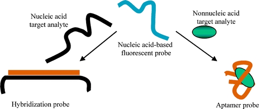
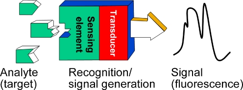
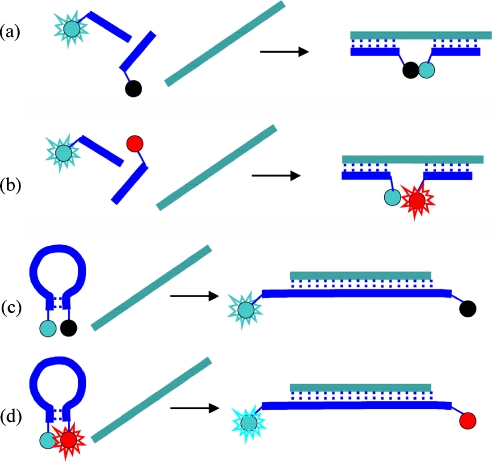
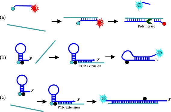
It is well known that nucleic acids play an essential role in living organisms because they store and transmit genetic information and use that information to direct the synthesis of proteins. However, less is known about the ability of nucleic acids to bind specific ligands and the application of oligonucleotides as molecular probes or biosensors. Oligonucleotide probes are single-stranded nucleic acid fragments that can be tailored to have high specificity and affinity for different targets including nucleic acids, proteins, small molecules, and ions. One can divide oligonucleotide-based probes into two main categories: hybridization probes that are based on the formation of complementary base-pairs, and aptamer probes that exploit selective recognition of nonnucleic acid analytes and may be compared with immunosensors. Design and construction of hybridization and aptamer probes are similar. Typically, oligonucleotide (DNA, RNA) with predefined base sequence and length is modified by covalent attachment of reporter groups (one or more fluorophores in fluorescence-based probes). The fluorescent labels act as transducers that transform biorecognition (hybridization, ligand binding) into a fluorescence signal. Fluorescent labels have several advantages, for example high sensitivity and multiple transduction approaches (fluorescence quenching or enhancement, fluorescence anisotropy, fluorescence lifetime, fluorescence resonance energy transfer (FRET), and excimer-monomer light switching). These multiple signaling options combined with the design flexibility of the recognition element (DNA, RNA, PNA, LNA) and various labeling strategies contribute to development of numerous selective and sensitive bioassays. This review covers fundamentals of the design and engineering of oligonucleotide probes, describes typical construction approaches, and discusses examples of probes used both in hybridization studies and in aptamer-based assays.
Figures
References
Publication types
MeSH terms
Substances
LinkOut - more resources
Full Text Sources
Other Literature Sources
