

Diffusion tensor MR imaging (DTI) metrics in the cervical spinal cord in asymptomatic HIV-positive patients
- PMID: 21046094
- PMCID: PMC3139090
- DOI: 10.1007/s00234-010-0782-6
Diffusion tensor MR imaging (DTI) metrics in the cervical spinal cord in asymptomatic HIV-positive patients
Abstract
Introduction: This study was conducted to compare diffusion tensor MR imaging (DTI) metrics of the cervical spinal cord in asymptomatic human immunodeficiency virus (HIV)-positive patients with those measured in healthy volunteers, and to assess whether DTI is a valuable diagnostic tool in the early detection of HIV-associated myelopathy (HIVM).
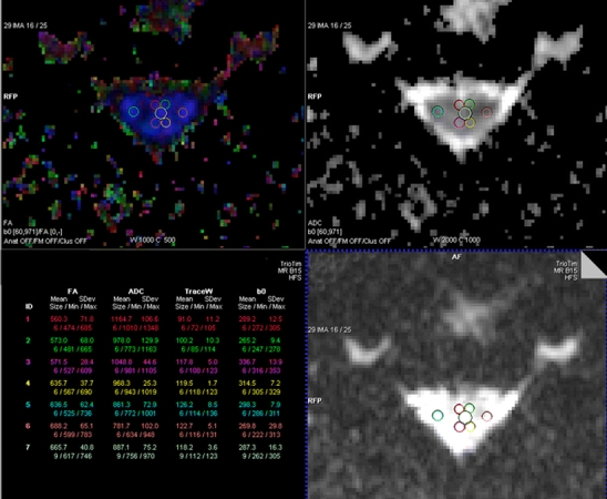
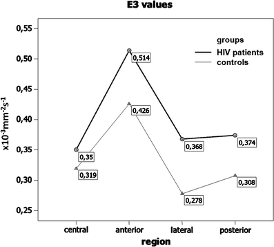
Methods: MR imaging of the cervical spinal cord was performed in 20 asymptomatic HIV-positive patients and in 20 healthy volunteers on a 3-T MR scanner. Average fractional anisotropy (FA), mean diffusivity (MD), and major (E1) and minor (E2, E3) eigenvalues were calculated within regions of interest (ROIs) at the C2/3 level (central and bilateral anterior, lateral and posterior white matter).
Results: Statistical analysis showed significant differences with regard to mean E3 values between patients and controls (p = 0.045; mixed-model analysis of variance (ANOVA) test). Mean FA was lower, and mean MD, mean E1, and mean E2 were higher in each measured ROI in patients compared to controls, but these differences were not statistically significant.
Conclusion: Asymptomatic HIV-positive patients demonstrate only subtle changes in DTI metrics measured in the cervical spinal cord compared to healthy volunteers that currently do not support using DTI as a diagnostic tool for the early detection of HIVM.
Figures
References
-
- Dal Pan GJ, Glass JD, McArthur JC. Clinicopathologic correlations of HIV-1-associated vacuolar myelopathy: an autopsy-based case-control study. Neurology. 1994;44:2159–2164. - PubMed
MeSH terms
LinkOut - more resources
Full Text Sources
Medical
Miscellaneous
