Case Reports
. 2011 Mar;5(1):92-9.
doi: 10.1007/s12105-010-0218-y. Epub 2010 Nov 3.

The spectrum of gnathic osteosarcoma: caveats for the clinician and the pathologist

Affiliations
Case Reports

The spectrum of gnathic osteosarcoma: caveats for the clinician and the pathologist

Ricardo J Padilla et al. Head Neck Pathol. 2011 Mar.

Abstract

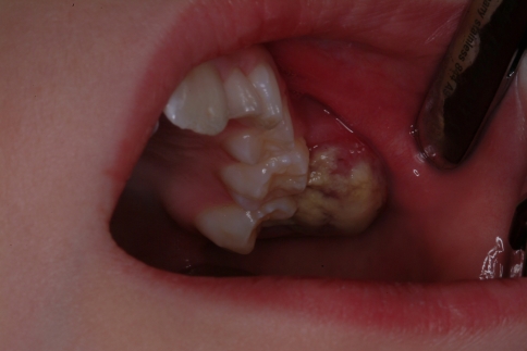
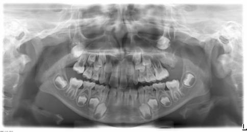
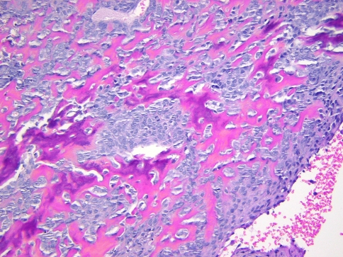
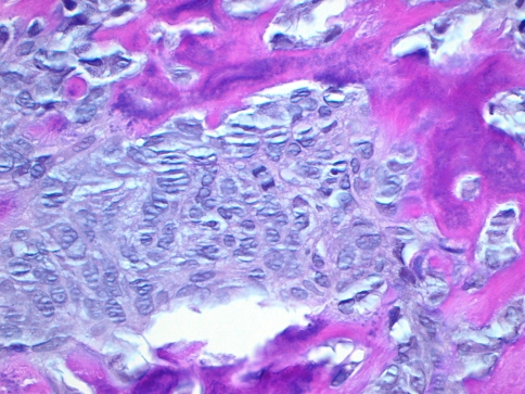
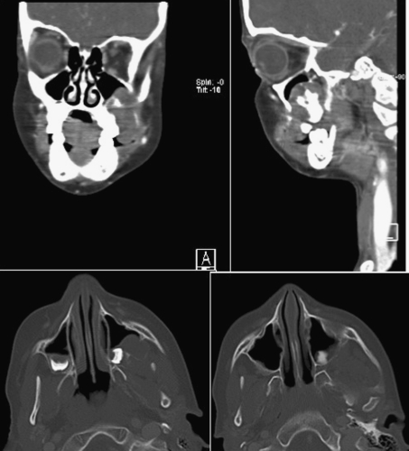
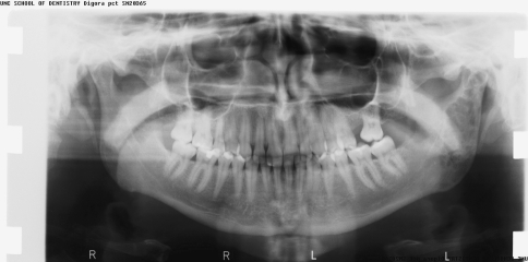
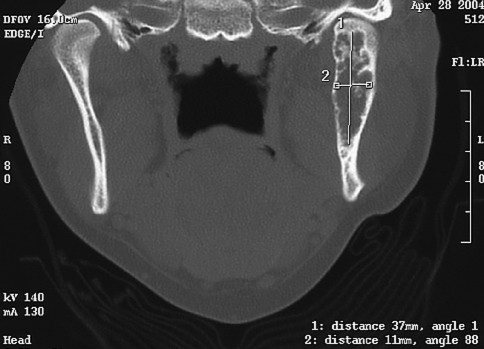
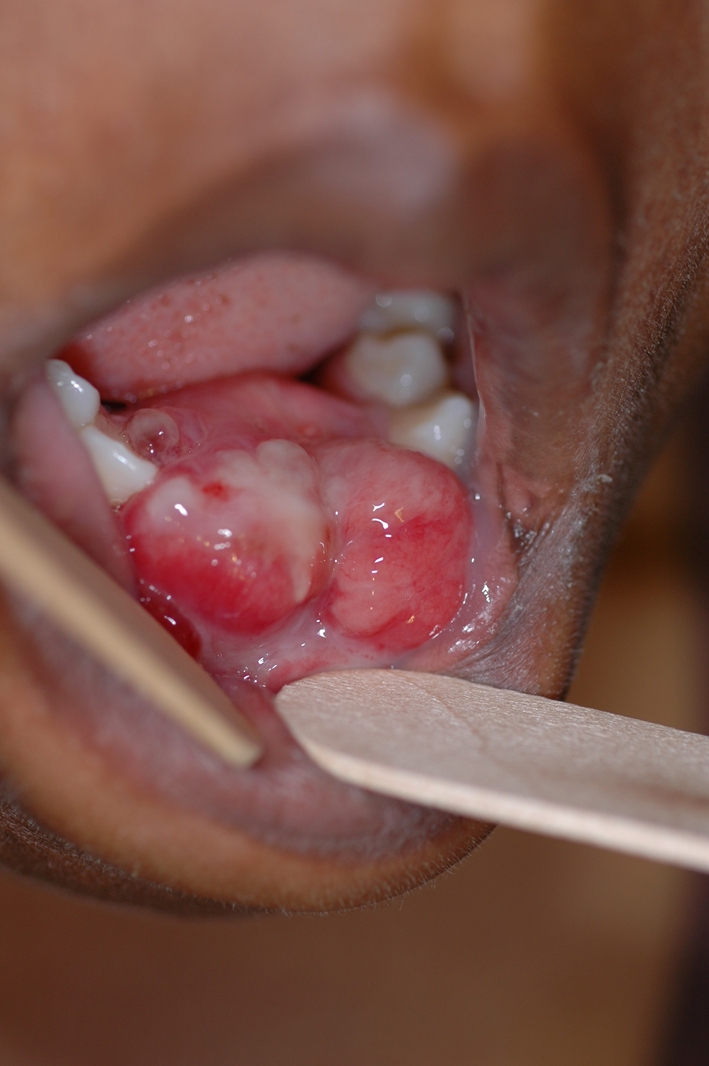
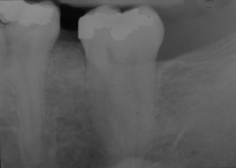
Seven expansile jaw lesions in patients ranging from 7 to 63 years are presented to illustrate diagnostic and management issues pertaining to cases ultimately proven to be gnathic osteosarcoma (GO). Six of the cases in our series were low-grade osteoblastic and one high-grade chondroblastic. None of our cases exhibited the characteristic "sunburst" radiopaque appearance described for GO. All of our cases displayed cortical expansion and one showed development of diastema. Two occurred in the maxilla and five in the mandible. Two of the patients with mandibular lesions presented initially with pain; all other patients were asymptomatic. Lack of pain resulted in a delay in diagnosis due to postponement of consultation or biopsy. Two cases underwent initial shallow non-representative biopsies, requiring a second biopsy for definitive diagnosis, further delaying treatment. Those biopsies were initially interpreted as pyogenic granuloma and peripheral ossifying fibroma, respectively. GO should always be considered in the differential diagnosis of expansile jaw lesions. Bone biopsies of lesions exhibiting pain and expansion of cortical plates should include medullary bone in order to minimize sampling error. In addition, all rapidly growing or painful exophytic bone lesions, and presumed soft tissue lesions that may involve underlying bone, should be examined histopathologically, and receive clinical and radiographic follow-up until complete resolution or healing is evident, regardless of the diagnosis. Based on the positive outcomes of the patients in our series, the prognosis of GO appears to be relatively favorable when compared to other sarcomas and osteosarcomas of long bones.


Figures

Fig. 1
Fig. 1
A 7-year-old boy (case # 1) with mass of the upper left maxilla
Fig. 2
Fig. 2
A panoramic radiograph of case #1 showing distal periodontal defect of tooth #14 with displaced impacted tooth # 15
Fig. 3
Fig. 3
Photomicrograph of the biopsy specimen from case # 1 exhibiting hypercellular stroma with mild cellular atypia and production of osteoid and bone. (Hematoxylin and eosin stain, 200× magnification)
Fig. 4
Fig. 4
Photomicrograph of case # 1 exhibiting atypical nuclear features including increased nuclear/cytoplasmic ratio, pleomorphism, and irregular chromatin distribution. Note also the production of osteoid by the malignant mesenchymal stroma. (Hematoxylin and eosin 400× magnification)
Fig. 5
Fig. 5
CT Scan of the maxillary region of patient in case # 1 showing the extent of the tumor. No other lesions were found elsewhere
Fig. 6
Fig. 6
Panoramic radiograph of lesion on case # 2. The lesion is present at mandibular angle, ramus, and condyle
Fig. 7
Fig. 7
Scan of the mandible of case # 2 showing expansion of the cortical plates over the radiolucent lesion
Fig. 8
Fig. 8
Large polypoid mass of anterior mandible (case #3) that grew to this size in 2 weeks
Fig. 9
Fig. 9
Bone scan (case #3) revealing a single skeletal active site located on the patient’s anterior mandible
Fig. 10
Fig. 10
Periapical radiograph of the area before development of the lesion from case # 4
Fig. 11
Fig. 11
Periapical radiograph of case # 6 showing a destructive lesion of the anterior maxilla at the time of second biopsy and diagnosis of GO
Fig. 12
Fig. 12
Periapical radiograph of case # 6 taken 18 months before the diagnosis of GO, which was submitted with the second biopsy, illustrated a diastema between 30 and 31 and increased alveolar bone production above the height of the adjacent alveolar crestal bone
Fig. 13
Fig. 13
Previous periapical radiograph of patient in case # 6 before the appearance of any lesion

Similar articles

Cited by

References

    1. Fletcher CDM, Unni KK, Mertens F, editors. World Health Organization classification of tumors. Pathology and genetics of tumors of soft tissue and bone. Lyon: IARC Press; 2002.
    1. Barnes L, Eveson JW, Reichart P, Sidransky D, editors. World Health Organization classification of tumours. Pathology and genetics of head and neck tumours. Lyon: IARC Press; 2005.
    1. Fernandez R, Nikitakis NG, Pazoli A, Ord RA. Osteogenic sarcoma of the jaw: a 10-year experience. J Oral Maxillofac Surg. 2007;65(7):1286–1291. doi: 10.1016/j.joms.2006.10.030. - DOI - PubMed
    1. Cheng YS, Wright JM, Walstad WR, Finn MD. Osteosarcoma arising in Paget’s disease of the mandible. Oral Oncol. 2002;38(8):785–792. doi: 10.1016/S1368-8375(02)00065-9. - DOI - PubMed
    1. Bramer JA, Abdulu AA, Tillman RM, Carter SR, Sumathi VP, Grimer RJ. Pre- and post-chemotherapy alkaline phosphatase levels as prognostic indicators in adults with localised osteosarcoma. Eur J Cancer. 2005;41(18):2846–2852. doi: 10.1016/j.ejca.2005.07.024. - DOI - PubMed

Publication types

LinkOut - more resources