

Intravesicular factors controlling exocytosis in chromaffin cells
- PMID: 21046452
- PMCID: PMC11498768
- DOI: 10.1007/s10571-010-9589-6
Intravesicular factors controlling exocytosis in chromaffin cells
Abstract
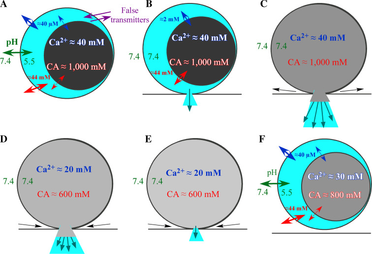
Chromaffin granules are similar organelles to the large dense core vesicles (LDCV) present in many secretory cell types including neurons. LDCV accumulate solutes at high concentrations (catecholamines, 0.5-1 M; ATP, 120-300 mM; or Ca(2+), 40 mM (Bulenda and Gratzl Biochemistry 24:7760-7765, 1985). Solutes seem to aggregate to a condensed matrix to elude osmotic lysis. The affinity of solutes for LDCV matrix is responsible for the delayed release of catecholamines during exocytosis. The aggregation of solutes occurs due to a specific H(+) pump denominated V-ATPase that maintains an inner acidic media (pH ≈5.5). This pH gradient against cytosol is also responsible for the vesicular accumulation of amines and Ca(2+). When this gradient is reduced by modulation of the V-ATPase activity, catecholamines and Ca(2+) are moved toward the cytosol. In addition, some drugs largely accumulate inside LDCV and not only impair the accumulation of natural solutes, but also act as false neurotransmitters when they are co-released with catecholamines. There is much experimental evidence to conclude that the physiological modulation of vesicle pH and the manipulation of intravesicular media with drugs affect the LDCV cargo and change the kinetics of exocytosis. Here, we will present some experimental data demonstrating the participation of drugs in the kinetics of exocytosis through changes in the composition of vesicular media. We also offer a model to explain the regulation of exocytosis by the intravesicular media that conciliate the experimentally obtained data.
Figures
Similar articles
-
Chromogranins A and B as regulators of vesicle cargo and exocytosis.Cell Mol Neurobiol. 2010 Nov;30(8):1181-7. doi: 10.1007/s10571-010-9584-y. Epub 2010 Nov 3. Cell Mol Neurobiol. 2010. PMID: 21046455 Free PMC article. Review.
-
Chromogranins as regulators of exocytosis.J Neurochem. 2010 Jul;114(2):335-43. doi: 10.1111/j.1471-4159.2010.06786.x. Epub 2010 Apr 29. J Neurochem. 2010. PMID: 20456013 Review.
-
Vesicular Ca(2+) mediates granule motion and exocytosis.Cell Calcium. 2012 Mar-Apr;51(3-4):338-41. doi: 10.1016/j.ceca.2011.12.009. Epub 2012 Jan 4. Cell Calcium. 2012. PMID: 22222091 Review.
-
How intravesicular composition affects exocytosis.Pflugers Arch. 2018 Jan;470(1):135-141. doi: 10.1007/s00424-017-2035-6. Epub 2017 Aug 4. Pflugers Arch. 2018. PMID: 28779472 Review.
-
The role of chromogranins in the secretory pathway.Biomol Concepts. 2013 Dec;4(6):605-9. doi: 10.1515/bmc-2013-0020. Biomol Concepts. 2013. PMID: 25436760 Review.
Cited by
-
Mice overexpressing chromogranin A display hypergranulogenic adrenal glands with attenuated ATP levels contributing to the hypertensive phenotype.J Hypertens. 2018 May;36(5):1115-1128. doi: 10.1097/HJH.0000000000001678. J Hypertens. 2018. PMID: 29389743 Free PMC article.
-
Amperometry methods for monitoring vesicular quantal size and regulation of exocytosis release.Pflugers Arch. 2018 Jan;470(1):125-134. doi: 10.1007/s00424-017-2069-9. Epub 2017 Sep 27. Pflugers Arch. 2018. PMID: 28951968 Free PMC article. Review.
-
Selective catecholamine recognition with NeuroSensor 521: a fluorescent sensor for the visualization of norepinephrine in fixed and live cells.ACS Chem Neurosci. 2013 Jun 19;4(6):918-23. doi: 10.1021/cn300227m. Epub 2013 Mar 25. ACS Chem Neurosci. 2013. PMID: 23527575 Free PMC article.
-
Multielectrode Arrays as a Means to Study Exocytosis in Human Platelets.Biosensors (Basel). 2023 Jan 4;13(1):86. doi: 10.3390/bios13010086. Biosensors (Basel). 2023. PMID: 36671921 Free PMC article.
-
Low pHo boosts burst firing and catecholamine release by blocking TASK-1 and BK channels while preserving Cav1 channels in mouse chromaffin cells.J Physiol. 2017 Apr 15;595(8):2587-2609. doi: 10.1113/JP273735. Epub 2017 Mar 2. J Physiol. 2017. PMID: 28026020 Free PMC article.
References
-
- Albillos A, Abad F, Garcia AG (1992) Cross-talk between M2 muscarinic and D1 dopamine receptors in the cat adrenal medulla. Biochem Biophys Res Commun 183:1019–1024 - PubMed
-
- Albillos A, Dernick G, Horstmann H, Almers W, Alvarez de Toledo G, Lindau M (1997) The exocytotic event in chromaffin cells revealed by patch amperometry. Nature 389:509–512 - PubMed
-
- Ales E, Tabares L, Poyato JM, Valero V, Lindau M, Alvarez de Toledo G (1999) High calcium concentrations shift the mode of exocytosis to the kiss-and-run mechanism. Nat Cell Biol 1:40–44 - PubMed
-
- Alvarez de Toledo G, Fernandez-Chacon R, Fernandez JM (1993) Release of secretory products during transient vesicle fusion. Nature 363:554–558 - PubMed
-
- Amatore C, Bouret Y, Travis ER, Wightman RM (2000) Adrenaline release by chromaffin cells: constrained swelling of the vesicle matrix leads to full fusion. Angew Chem Int Ed 39:1952–1955 - PubMed
MeSH terms
Substances
LinkOut - more resources
Full Text Sources
Miscellaneous
