. 2011 Feb;39(2):674-87.
doi: 10.1007/s10439-010-0197-x. Epub 2010 Oct 29.

Non-thermal plasma induces apoptosis in melanoma cells via production of intracellular reactive oxygen species

Affiliations

Non-thermal plasma induces apoptosis in melanoma cells via production of intracellular reactive oxygen species

Rachel Sensenig et al. Ann Biomed Eng. 2011 Feb.

Retraction in

Abstract

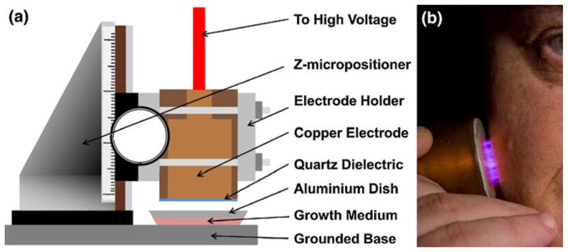
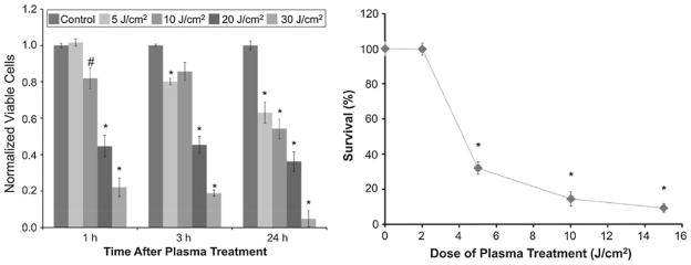
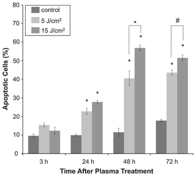
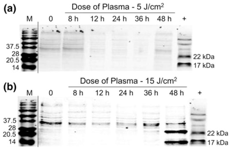
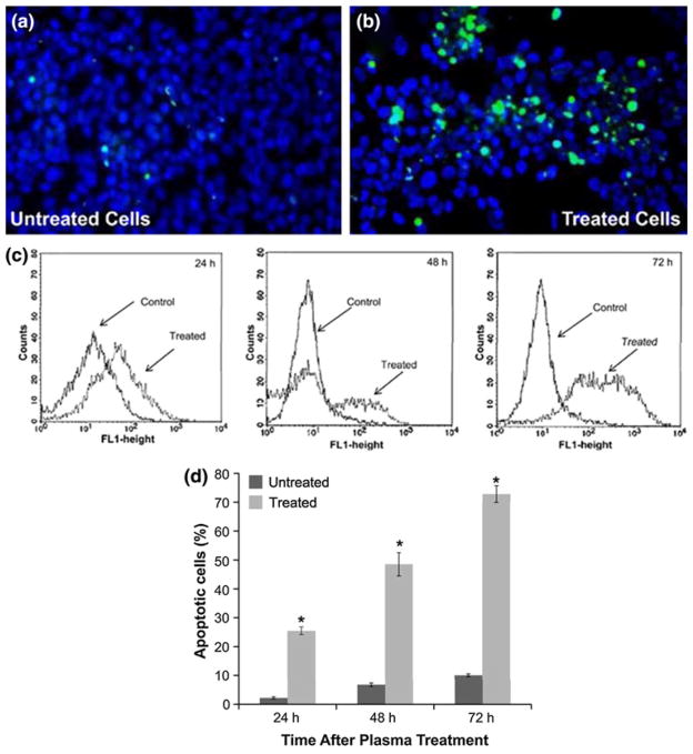
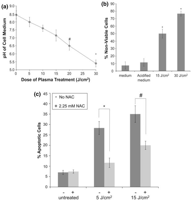
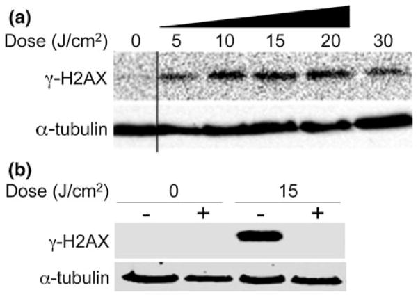
Non-thermal atmospheric pressure dielectric barrier discharge (DBD) plasma may provide a novel approach to treat malignancies via induction of apoptosis. The purpose of this study was to evaluate the potential of DBD plasma to induce apoptosis in melanoma cells. Melanoma cells were exposed to plasma at doses that did not induce necrosis, and cell viability and apoptotic activity were evaluated by Trypan blue exclusion test, Annexin-V/PI staining, caspase-3 cleavage, and TUNEL® analysis. Trypan blue staining revealed that non-thermal plasma treatment significantly decreased the viability of cells in a dose-dependent manner 3 and 24 h after plasma treatment. Annexin-V/PI staining revealed a significant increase in apoptosis in plasma-treated cells at 24, 48, and 72 h post-treatment (p < 0.001). Caspase-3 cleavage was observed 48 h post-plasma treatment at a dose of 15 J/cm(2). TUNEL® analysis of plasma-treated cells demonstrated an increase in apoptosis at 48 and 72 h post-treatment (p < 0.001) at a dose of 15 J/cm(2). Pre-treatment with N-acetyl-L: -cysteine (NAC), an intracellular reactive oxygen species (ROS) scavenger, significantly decreased apoptosis in plasma-treated cells at 5 and 15 J/cm(2). Plasma treatment induces apoptosis in melanoma cells through a pathway that appears to be dependent on production of intracellular ROS. DBD plasma production of intracellular ROS leads to dose-dependent DNA damage in melanoma cells, detected by γ-H2AX, which was completely abrogated by pre-treating cells with ROS scavenger, NAC. Plasma-induced DNA damage in turn may lead to the observed plasma-induced apoptosis. Since plasma is non-thermal, it may be used to selectively treat malignancies.


Figures

FIGURE 1
FIGURE 1
Non-thermal plasma treatment of melanoma cells. (a) Schematic of the non-thermal atmospheric pressure DBD plasma treatment setup showing the high voltage electrode and the sample holder. Note: The depth of medium covering the cells has been exaggerated for visual clarity, but the actual depth is a few 100 μm. (b) Non-thermal atmospheric pressure DBD plasma applied safely to human tissue.
FIGURE 2
FIGURE 2
Growth rates of melanoma cells in poly L-lysine coated dishes and plastic petri dishes. 1 × 10⁶ cells were seeded at day 0 in aluminum and plastic petri dishes. Viability was evaluated using trypan blue exclusion test each day for 12 days after seeding. Data from triplicate samples (±SD) were plotted. Growth was monitored by replacing medium every 2 days.
FIGURE 3
FIGURE 3
Non-thermal DBD plasma treatment leads to dose-dependent decrease in viability and survivability of melanoma cells. (a) Melanoma cells were exposed to non-thermal DBD plasma at the indicated doses. Cell viability was analyzed 1, 3, and 24 h after plasma treatment by trypan blue exclusion test. Briefly, floating and trypsin-detached cells were centrifuged and resuspended in 2-mL PBS. 20-μL cell suspension was mixed with 20-μL trypan blue and incubated for 5 min at room temperature. Viable and non-viable cells were counted on a hemocytometer. Data from triplicate samples (±SD) are expressed relative to total number of cells in untreated control. (b) Cells were treated with the indicated dose of DBD plasma; and colony survival assays were performed as described. Data from triplicate samples (±SD) are expressed relative to the number of colonies in the untreated control.
FIGURE 4
FIGURE 4
Early apoptotic activity in melanoma cells after non-thermal plasma treatment is confirmed by Annexin-V/PI staining. Apoptotic activity in plasma-treated melanoma cells was analyzed 3, 24, and 72 h after treatment with the indicated dose of DBD plasma, by harvesting and staining cells with Annexin-V/propidium iodide (PI) according to the manufacturer’s protocol. Samples were run on Guava® flow cytometer and analyzed by Guava® flow cytometry software. Data from triplicate samples (±SD) are plotted.
FIGURE 5
FIGURE 5
Caspase-3 cleavage assay confirms that nonthermal plasma treatment initiates apoptosis in melanoma cells. Melanoma cells were treated with non-thermal plasma at two different doses, (a) 5 J/cm2 and (b) 15 J/cm2. Caspase-3 cleavage was analyzed by preparing lysates 8, 12, 24, 36, and 48 h after plasma treatment by immunoblot. Representative immunoblots with antibody to cleaved caspase-3 are shown.
FIGURE 6
FIGURE 6
TUNEL® assay demonstrates that non-thermal plasma treatment induces late-stage apoptosis in melanoma cells. Images of (a) untreated and (b) treated melanoma cells following TUNEL assay. All cells are stained blue (darker circles) and apoptotic cells are also stained green (bright spots). Assay performed 24 h following treatment at a dose of 15 J/cm2. (c) The FL1 (fluorescein) fluorescence histogram (number of events over fluorescence intensity) of a representative experiment is shown. (d) Flow cytometry was performed as described and percentage of TUNEL-positive cells from triplicate samples (±SD) in untreated and treated (15 J/cm2) samples analyzed via flow cytometry 24, 48, and 72 h after plasma treatment are plotted.
FIGURE 7
FIGURE 7
pH does not play a major role in plasma-induced apoptosis, while plasma-generated intracellular ROS mediate induction of apoptosis in DBD plasma-treated melanoma cells. (a) pH of the medium covering the cells during treatment was measured using a micro pH electrode after treatment with different doses of plasma as indicated. Data from triplicate samples (±SD) are plotted. (b) Melanoma cells were exposed to fresh cell growth medium or acidified cell growth medium with a pH of 5.4 for 1 min or plasma treated at a dose of 30 J/cm2 followed by incubation in fresh medium for 3 h. Cell viability (non-viable cells stained blue) was determined 3 h after treatment by trypan blue exclusion test. Data from triplicate samples (±SD) are plotted. (c) Melanoma cells were incubated for 1 h with 2.25 mM N-acetyl-L-cysteine (NAC) (+) or cell culture medium (−), followed by treatment with the indicated dose of DBD plasma. Apoptotic activity was measured 24 h after plasma treatment by harvesting and staining the cells with Annexin-V/PI. Samples were run on the flow cytometer and analyzed using Gauva® software. Data from triplicate samples (±SD) are plotted. * p <0.001 while # p<0.05.
FIGURE 8
FIGURE 8
Intracellular ROS mediates induction of DNA damage by DBD plasma. (a) Melanoma cells were treated with the indicated dose of plasma as described. After 1-h incubation, lysates were prepared and resolved by SDS-PAGE, and representative immunoblots with antibody to γ-H2AX (top) or α-tubulin (bottom) are shown. (b) Melanoma cells were incubated for 2 h with 4 mM N-acetyl-L-cysteine (NAC) (+) or cell culture medium (−), followed by treatment with the indicated dose of DBD plasma. γ-H2AX (top) or α-tubulin (bottom) was detected by immunoblot of cell lysates prepared 1-h after plasma treatment.

References

    1. Berk LB. Radiation therapy as primary and adjuvant treatment for local and regional melanoma. Cancer Control. 2008;15(3):233–238. - PubMed
    1. Colt HG, Crawford SW. In vitro study of the safety limits of bronchoscopic argon plasma coagulation in the presence of airway stents. Respirology. 2006;11(5):643–647. - PubMed
    1. Coulombe S. Live cell permeabilization using the APGD-t. 1st International Conference on Plasma Medicine (ICPM); TX: Corpus Christi; 2007.
    1. Coulombe S, et al. Miniature atmospheric pressure glow discharge torch (APGD-t) for local biomedical applications. Pure Appl Chem. 2006;78(6):1137–1146.
    1. Eliasson B, Egli W, Kogelschatz U. Modelling of dielectric barrier discharge chemistry. Pure Appl Chem. 1994;66(6):1275–1286.

Publication types