

Review
doi: 10.1021/mp100225y.
Epub 2010 Dec 17.
The art of engineering viral nanoparticles
Affiliations
- PMID: 21047140
- PMCID: PMC3156490
- DOI: 10.1021/mp100225y
Item in Clipboard
Review
The art of engineering viral nanoparticles
Mol Pharm.
.
Abstract
Viral nanotechnology is an emerging and highly interdisciplinary field in which viral nanoparticles (VNPs) are applied in diverse areas such as electronics, energy and next-generation medical devices. VNPs have been developed as candidates for novel materials, and are often described as "programmable" because they can be modified and functionalized using a number of techniques. In this review, we discuss the concepts and methods that allow VNPs to be engineered, including (i) bioconjugation chemistries, (ii) encapsulation techniques, (iii) mineralization strategies, and (iv) film and hydrogel development. With all these techniques in hand, the potential applications of VNPs are limited only by the imagination.
Figures
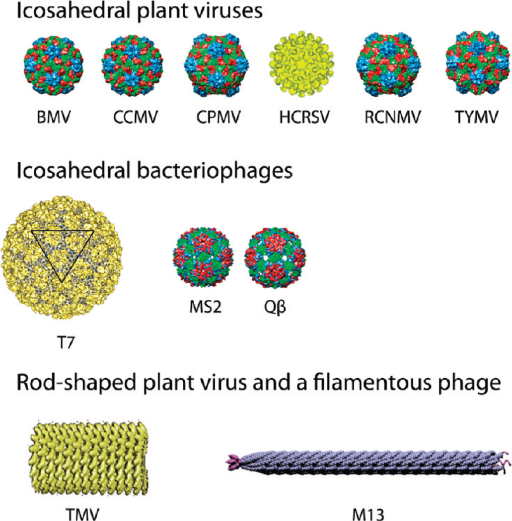
An overview of the viral nanoparticles (VNPs) that have been developed for materials science and medicine. Icosahedral plant viruses: Brome mosaic virus (BMV), Cowpea cholorotic mottle virus (CCMV), Cowpea mosaic virus (CPMV), Hibiscus cholorotic ringspot virus (HCRSV), Red clover necrotic mottle virus (RCNMV), Turnip yellow mosaic virus (TYMV). Icosahedral bacteriophages: T7, MS2, and Qβ. Note that T7 is a head–tail phage, but only the head is shown. Rod-shaped and filamentous viruses: Tobacco mosaic virus (TMV) and phage M13. Images of the following VNPs were reproduced with permission from the VIPER database (www.viperdb.scripps.edu ; Carrillo-Tripp, M.; Shepherd, C. M.; Borelli, I. A.; Venkataraman, S.; Lander, G.; Natarajan, P.; Johnson, J. E.; Brooks, C. L., III; Reddy, V. S. VIPERdb2: an enhanced and web API enabled relational database for structural virology. Nucleic Acids Res. 2009, 37, D436–D442. DOI: 10.1093/nar/gkn840): BMV, CCMV, CPMV, RCNMV, TYMV, MS2, Qβ. The structure of HCRSV was reproduced with permission from Doan, D. N., et al. J. Struct. Biol. 2003, 144 (3), 253–261. Copyright 2003 Elsevier. The T7 structure was reproduced with permission from Agirrezabala, X., et al. Structure 2007, 15, 461–472. Copyright 2007 Elsevier. TMV was reproduced with permission from ref . Copyright 2008 Annual Reviews. M13 was reproduced with permission from ref . Copyright 2007 National Academy of Sciences.
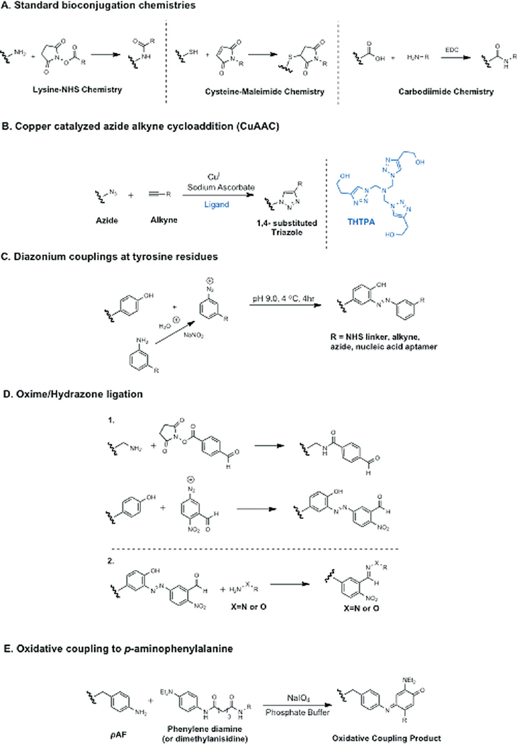
Bioconjugation methods for VNPs. (A) An overview of the three most common bioconjugation chemistries, typically addressing the most reactive amino acid residues and often carried out using commercially available reagents. (B) The copper-catalyzed azide alkyne cycloaddition reaction is a high-fidelity bioorthogonal reaction that can be carried out at low reagent concentrations. (C) Diazonium coupling chemistry is a more specialized and labor-intensive coupling process. Experienced chemists, however, should find this a versatile method for the introduction of new functional groups onto VNPs if standard bioconjugation protocols are inappropriate. (D) Condensation reactions to form oximes and hydrazones. (1) Two strategies for the introduction of aldehydes into proteins. (2) Bioorthogonal condensation reaction. (E) Oxidative coupling to the unnatural amino acid p-aminophenylalanine, a specific reaction that allows labeling of the unnatural amino acid in the presence of reactive side chains.
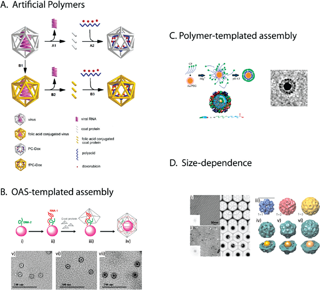
(A) Artificial polymers. Schematic illustration of the preparation of a doxorubicin-loaded HCSRV protein cage with and without folic acid conjugation (fPC-Dox, PC-Dox). Steps A1 and B2 indicate the removal of viral RNA from the plant virus and purification of coat proteins. Steps A2 and B3 involve the encapsulation of polyacid and doxorubicin during the reassembly of the protein cage. Step B1 refers to the conjugation of folic acid onto the viral coat protein. Reproduced with permission from ref . Copyright 2007 American Chemical Society. (B) OAS-templated assembly. Top panel shows the assembly of RCNMV VLPs around a gold nanoparticles by OAS templating. (i) Conjugation of nanoparticle with DNA-2; (ii) addition of RNA-1 interacts with DNA-2 to form the functional OAS; (iii) the artificial OAS then templates the assembly of coat protein; and (iv) formation of VLP with enclosed nanoparticle. Bottom panel shows negatively stained transmission electron microscopy images of VLP encapsulating (v) 4 nm CoFe2O4, (vi) 10 nm CoFe2O4, and (vii) 15 nm CoFe2O4 nanoparticles after purification. Reproduced with permission from ref . Copyright 2007 American Chemical Society. (C) Polymer-templated assembly. (i) Proposed mechanism of VLP assembly from coat proteins (CP). First, electrostatic interaction leads to the formation of disordered protein-gold nanoparticle complexes. The second step is a crystallization phase in which the protein–protein interactions lead to the formation of a regular capsid. Schematic depiction of the encapsidated nanoparticle functionalized with carboxyl-terminated tri(ethylene glycol) monomethyl ether (TEG) chains. Right panel: Cryo-electron micrograph of a single VLP. The regular arrangement of the protein structure coating the 12 nm diameter gold nanoparticle (black disk) is evident. The averages have been obtained by superposition of 10 individual images, in each case. Reproduced with permission from ref . Copyright 2006 American Chemical Society. (D) Size-dependence. Left panel: Negatively stained electron micrographs, Fourier transforms (insets), and corresponding Fourier projection maps. (i) BMV 2D crystal. The lattice constant is 26 nm (one unit cell is shown), and the arrangement of the densities suggests a T = 3 structure. (ii) VLPs containing 12 nm gold cores arranged in a 2D lattice. The lattice constant is 25 nm. Right panel: 3D reconstructions of BMV and VLP using negative stain data. (iii) T = 1, 2, and 3 models of BMV capsids. The T = 1 and pseudo T = 2 structures were obtained from the Virus Particle ExplorR (VIPER) database. The T = 3 structure is the reconstructed image of BMV in this work (scale bar, 21 nm.) (iv) VLPs containing 6 nm gold cores are characterized by the absence of electron density at the 3-fold symmetry axes. The structure and diameter is close to a T = 1 capsid. (v) The VLPs containing 9 nm gold cores are reminiscent of a pseudo T = 2 structure. The presence of electron density at the 3-fold axes distinguishes it from the VLP structure with 6 nm cores. (vi) The shape of the VLPs containing 12 nm gold cores resembles the spherical shape of BMV although it still lacks clear evidence of hexameric capsomers. Concentric layering is a characteristic of all VLPs. Reproduced with permission from ref . Copyright 2007 National Academy of Sciences.
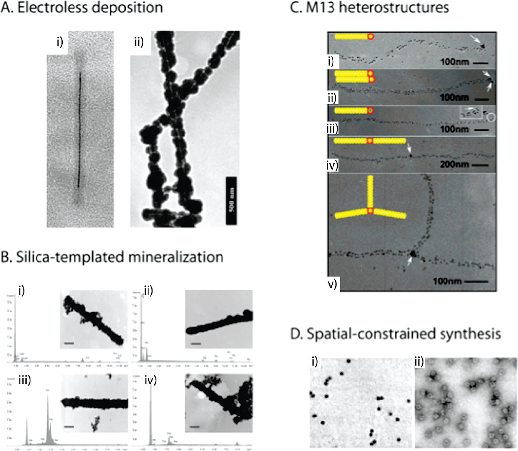
(A) Electroless deposition. Transmission electron micrographs of metalized TMV particles produced by electroless deposition. (i) TMV after Pd(II) activation, followed by electroless deposition of Ni. TMV is filled with a nickel wire with a ca. 3 nm diameter. Reproduced with permission from ref . Copyright 2003 American Chemical Society. (ii) TMV metalized with nickel on the external surface. Reproduced with permission from ref . Copyright 2004 Wiley. (B) Silica-templated mineralization. Transmission electron micrographs and accompanying energy dispersive X-ray spectra of (i) Au on thick-shell silica-coated TMV template, (ii) Ag on thick-shell silica-coated TMV template, (iii) Pt on thick-shell silica-coated TMV template, and (iv) Pd on thick-shell silica-coated TMV template. Al, Fe, and Cu peaks in the energy dispersive spectroscopy spectra are due to background effects. Scale bar = 100 nm. Reproduced with permission from 84. Copyright 2009 Elsevier. (C) M13 heterostructures. (i–v) Transmission electron micrographs of various nanoarchitectures templated by M13. Gold nanoparticles (~5 nm) bind to genetically engineered pVIII proteins along the virus axis and form 1D arrays, while a second peptide motif on pIII protein simultaneously binds to streptavidin-coated nanoparticles. Arrows highlight the streptavidin-conjugated gold nanoparticles (~15 nm) and CdSe quantum dots bound on pIII proteins. The insets show the assembly schemes of observed structures. White represents the virus structure, yellow dots represent gold nanoparticles, the green dot represents a CdSe quantum dot, and red represents the streptavidin coating around gold or CdSe particles. (C, inset) The enlarged image of the CdSe quantum dot attached to the end of the virus. Reproduced with permission from ref . Copyright 2005 American Chemical Society. (D) Spatially constrained synthesis. Transmission electron micrographs of paratungstate-mineralized CCMV particles after isolation by centrifugation on a sucrose gradient. (i) An unstained sample showing discrete electron dense cores; (ii) a negatively stained sample of A showing the mineral core surrounded by the intact virus protein cage. Reproduced with permission from ref . Copyright 1998 Nature Publishing Group.
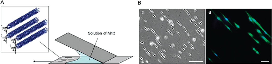
Strategy for aligned deposition of M13. (A) Aligned thin films of modified M13 particles were deposited onto glass surfaces by slowly dragging the meniscus of a high concentration solution across the surface. The shear force generated in the process caused the particles to align in the direction of the applied shear force. (B) Optical and fluorescent confocal micrographs of CHO cells cultured on aligned M13-RGD thin films. Reproduced with permission from ref . Copyright 2008 Royal Society of Chemistry.
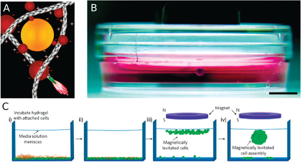
(A) Scheme of electrostatic interactions between magnetic iron oxide (MIO; brown spheres) and gold nanoparticles (yellow spheres) with phage (elongated structures; pIII and pVIII indicate surface capsid proteins). Nanoparticles are not drawn to scale. (B) Human glioblastoma cells (lower arrow) treated with MIO-containing hydrogel held at the air-medium interface by a magnet. The image was captured after 48 h in culture and depicts a ~1 mm spheroid. Scale bar = 5 mm. (C) (i) Hydrogel is dispersed over cells and the mixture is incubated. (ii) Washing steps remove non-interacting hydrogel fragments. Fractions of phage, gold and MIO nanoparticles enter cells or remain membrane-bound. (iii) An external magnet causes cells to rise to the air-medium interface. (iv) After 12 h of levitation, characteristic multicellular structures form (a single structure is shown here). Reproduced with permission from ref . Copyright 2010 Nature Publishing Group.
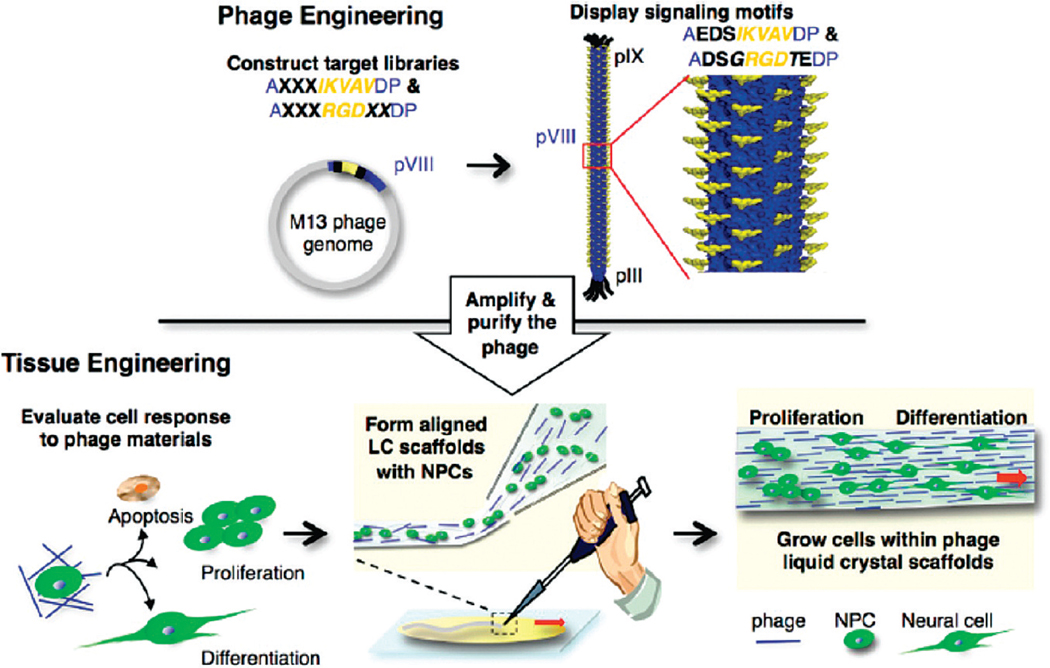
Schematic diagram of the M13 phage tissue engineering process used by the Lee group, showing phage engineering, cell response characterization, and the fabrication of an aligned fiber matrix. Reproduced with permission from ref . Copyright 2009 American Chemical Society.
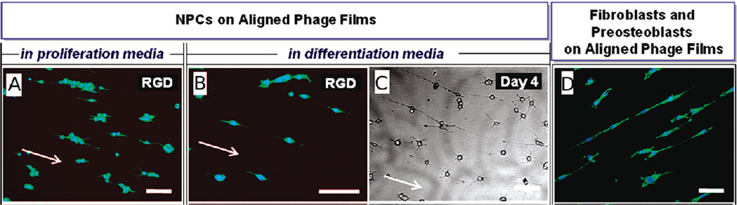
Growth of neuroprogenitor cells (NPCs) and fibroblasts on aligned M13 thin films. (A) Aligned RGD-phage film (day 1) in proliferation medium. (B) Aligned RGD-phage film (day 1) in differentiation medium. (C) Bright-field optical micrograph of NPCs on the aligned RGD-phage film at day 4. (D) Composite fluorescence images of NIH-3T3 fibroblasts grown on the aligned RGD-phage (12 h). Actin fibers and nuclei were stained with phalloidin and DAPI, respectively. Scale bars = 100 µm. Reproduced with permission from ref . Copyright 2010 American Chemical Society.
References
-
- Zaitlin M. The discovery of the causal agent of the tobacco mosaic disease. In: Kung S-D, Yang S-F, editors. Discoveries in Plant Biology. Hong Kong: World Publishing; 1998. pp. 105–110.
-
- Clark JR, March JB. Bacteriophages and biotechnology: vaccines, gene therapy and antibacterials. Trends Biotechnol. 2006;24(5):212–218. - PubMed
-
- Marks T, Sharp R. Bacteriophages and biotechnology: a review. J. Chem. Technol. Biotechnol. 2000;75:6–17.
-
- Garcea RL, Gissmann L. Virus-like particles as vaccines and vessels for the delivery of small molecules. Curr. Opin. Biotechnol. 2004;15(6):513–517. - PubMed
Publication types
MeSH terms
Grants and funding
LinkOut - more resources
Full Text Sources
Other Literature Sources
