. 2011 Jan;28(1):61-8.
doi: 10.1017/S0952523810000301. Epub 2010 Nov 4.

Nonlinearity and noise at the rod-rod bipolar cell synapse

Affiliations

Nonlinearity and noise at the rod-rod bipolar cell synapse

E Brady Trexler et al. Vis Neurosci. 2011 Jan.

Abstract

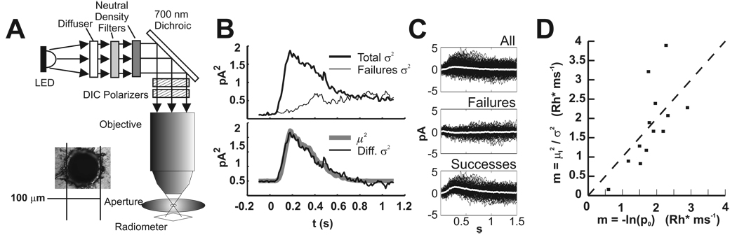
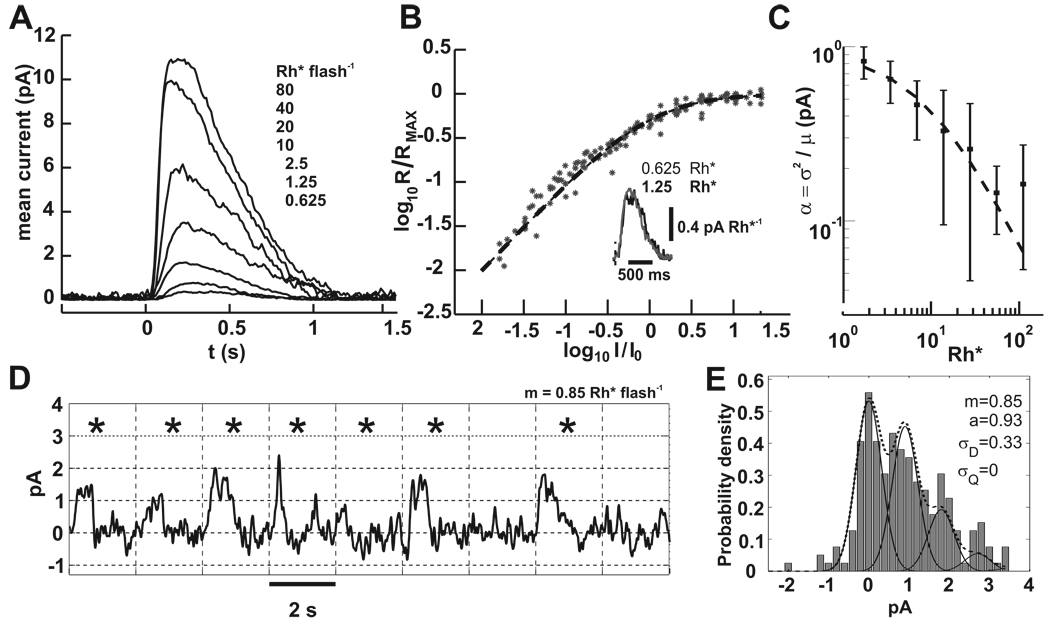
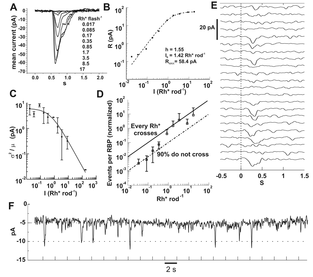
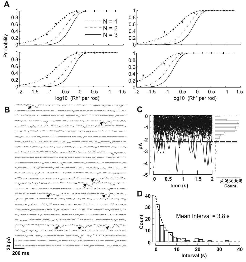
In the retina, rod bipolar (RBP) cells synapse with many rods, and suppression of rod outer segment and synaptic noise is necessary for their detection of rod single-photon responses (SPRs). Depending on the rods' signal-to-noise ratio (SNR), the suppression mechanism will likely eliminate some SPRs as well, resulting in decreased quantum efficiency. We examined this synapse in rabbit, where 100 rods converge onto each RBP. Suction electrode recordings showed that rabbit rod SPRs were difficult to distinguish from noise (independent SNR estimates were 2.3 and 2.8). Nonlinear transmission from rods to RBPs improved response detection (SNR = 8.7), but a large portion of the rod SPRs was discarded. For the dimmest flashes, the loss approached 90%. Despite the high rejection ratio, noise of two distinct types was apparent in the RBP traces: low-amplitude rumblings and discrete events that resembled the SPR. The SPR-like event frequency suggests that they result from thermal isomerizations of rhodopsin, which occurred at the rate 0.033/s/rod. The presence of low-amplitude noise is explained by a sigmoidal input-output relationship at the rod-RBP synapse and the input of noisy rods. The rabbit rod SNR and RBP quantum efficiency are the lowest yet reported, suggesting that the quantum efficiency of the rod-RBP synapse may depend on the SNR in rods. These results point to the possibility that fewer photoisomerizations are discarded for species such as primate, which has a higher rod SNR.


Figures

Figure 1
Figure 1. Calibration of light stimuli by rod suction recordings
A. Schematic of the optical path. Between the objective and the aperture is a water droplet. The distance from the aperture to the radiometer is ~5 mm. We believe this causes an error in quantitating photon flux due to improper focusing. Inset: a micrograph of the 100µ aperture used to limit area for flux calculation. B. Variance subtraction for determining µ22. The top panel shows the total variance and the variance of failures. Failures were chosen according to correlation of each trace with the mean. The bottom panel shows the overlay of the subtracted variance and the square of the mean. The ratio of the traces is one, indicating that the flash delivered, on average, 1 Rh*. C. Successes and failures chosen by correlation with the mean. Top panel shows all traces and the mean (white). Middle: correlation < 0.51 chooses failures. Bottom: correlation >0.51 chooses successes. D. Comparison of calibration methods. Each method gave 1.7 Rh* per ms.
Figure 2
Figure 2. Rod suction electrode recordings demonstrate the noise that obscures the SPR
A) A family of responses to increasing flash energies. Each trace is the average of 10 to 100 flash presentations. Flashes were delivered at 0.5 s. Dark current for this cell was ~12 pA and the intensity that produced the half maximal response was 17.25 Rh*. B) Data collected from 15 rods was fit to equation (3). Responses for each rod were normalized to the dark current and scaled by the intensity that produced the half maximal response. Inset: an overlay of the two lowest flash strengths divided by their flash energies shows the similarity in waveform, which is expected for linear summation. Flash onset is indicated by the vertical dashed line. C) The contribution of each Rh* to the flash response decreases with increasing stimulus strength. Plotted is the ratio of the variance to the mean at the peak of the flash response.. Data from 15 rods was fit to equation (4) with parameters I0 = 8.4 Rh* and a = 0.92 pA. D) A segment of a 400 s rod suction recording that encompassed 9 flash presentations at 0.5 Hz is shown. Flashes delivered on average 0.85 Rh*, and from Poisson statistics ~40% should have generated no response. Tick marks on the abscissa and vertical grid lines correspond to the flash times. Asterisks mark the flash responses whose correlation with the mean response was > 0.5. From Poisson statistics, singletons and multiples should have occured with probabilities 0.36 and 0.21, respectively. Baseline drift was corrected by fitting and then subtracting a spline to the average current of 100 ms periods in darkness that were spaced 2 s apart. Bandwidth = DC to 5 Hz. E) The response amplitude histogram of the entire 400 s record in D is shown (200 flashes). No spline subtraction was used. Amplitudes were generated by correlation with a scaled portion of the mean flash response with each trial (Field and Rieke, 2002). The dotted line is the from equation (5), with individual peaks corresponding to 0, 1, 2, and 3 photoisomerizations shown by the solid lines. Only a and σD parameters were allowed to vary. Best fit parameters are shown.
Figure 3
Figure 3. RBP responses depend nonlinearly on flash strength
A) Baseline subtracted currents recorded from a RBP in the perforated patch configuration. Each trace is an average of 3 to 20 flash presentations. Flashes were delivered at 0.5 s. Vh = −60 mV. B) Peak currents of A plotted against flash intensity were fitted to equation (3). Best fit parameters are displayed in the plot. C) The ratio of the ensemble variance to the mean yielded the unitary response amplitude for RBPs (n=12). As flash intensity increases, the current contributed by each Rh* decreases. The solid line is equation (4) with parameters aRBP = 6.1 pA, I0 = 0.52 Rh* rod−1. D) Rh* events in rods are discarded by RBPs. The number of unitary events in a RBP is given by equation (2). Data from individual cells (n=5) was averaged and scaled so that the points above 1 Rh* were bounded by the upper black line, which marks the input-output relationship corresponding to synaptic transfer of every Rh* in the rods. The lower line was drawn to mark 90% loss due to nonlinear transfer. The transition from 10% to 100% transmission occurs over the range of 0.3 to 2 Rh*, in which the probablility of multiple photon absorptions and the consequent threshold crossings also increases. E) An example of a RBP response to repeated flashes delivering on average 0.17 Rh* rod−1. Note the low amplitude noise during periods without a stimulus. F) Same data in E, but the trials are concatenated. The holding current for this cell was 5 pA. Most of the successful responses have amplitude of 5 or 6 pA, as suggested by the fit in C. Tick marks denote flash timing. Bandwidth = DC to 5 Hz for A, E and F.
Figure 4
Figure 4. RBPs respond to single photon absorptions and noise in RBPs may result from thermal isomerizations of rhodopsin
A) Examples of fits for 4 RBPs to cumulative Poisson distributions for the requirements of 1, 2 or 3 photons (N). Despite the steep nonlinearity at the rod-RBP synapse, RBPs respond to single photons. B) RBPs in darkness exhibit low amplitude as well as larger discrete noise events. A 56 s portion of a 180 s recording of a RBP in darkness is shown. The current was segmented into 2 s sweeps for display purposes. Bandwidth = DC to 5 Hz. There are several discrete events that resemble the single photon responses depicted in Figure 3E and F. Events that exceed the threshold in C are marked with arrows. C) Ninety two-second sweeps from the cell in A were match filtered to isolate events resembling the time course of the single photon response and overlaid. The dashed line indicates placement of the event detection threshold, which was set at the mean plus 3 times the standard deviation of the entire record. The histogram to the right shows the amplitudes of all downward deflections in the current. Most of the low amplitude noise appears normally distributed, but not the events above threshold, which we consider the result of thermal isomerizations. D) Intervals between events detected by threshold crossings were binned and fit to an exponential to find the mean interval. The plotted histogram was generated from data compiled from 8 RBP cells, providing an estimate of 3.8 s between SPR-like events in RBPs.

Similar articles

Cited by

References

    1. Barlow HB. Increment thresholds at low intensities considered as signal/noise discriminations. J Physiol. 1957;136(3):469–488. - PMC - PubMed
    1. Baylor DA, Lamb TD, Yau KW. The membrane current of single rod outer segments. J Physiol. 1979a;288:589–611. - PMC - PubMed
    1. Baylor DA, Lamb TD, Yau KW. Responses of retinal rods to single photons. J Physiol. 1979b;288:613–634. - PMC - PubMed
    1. Baylor DA, Nunn BJ, Schnapf JL. The photocurrent, noise and spectral sensitivity of rods of the monkey macaca fascicularis. J Physiol. 1984;357:575–607. - PMC - PubMed
    1. Berntson A, Smith RG, Taylor WR. Transmission of single photon signals through a binary synapse in the mammalian retina. Vis Neurosci. 2004;21(5):693–702. - PubMed

Publication types