. 2011 Feb 10;149(3):323-31.
doi: 10.1016/j.jconrel.2010.10.031. Epub 2010 Nov 1.

Enhanced endothelial delivery and biochemical effects of α-galactosidase by ICAM-1-targeted nanocarriers for Fabry disease

Affiliations

Enhanced endothelial delivery and biochemical effects of α-galactosidase by ICAM-1-targeted nanocarriers for Fabry disease

Janet Hsu et al. J Control Release. .

Abstract

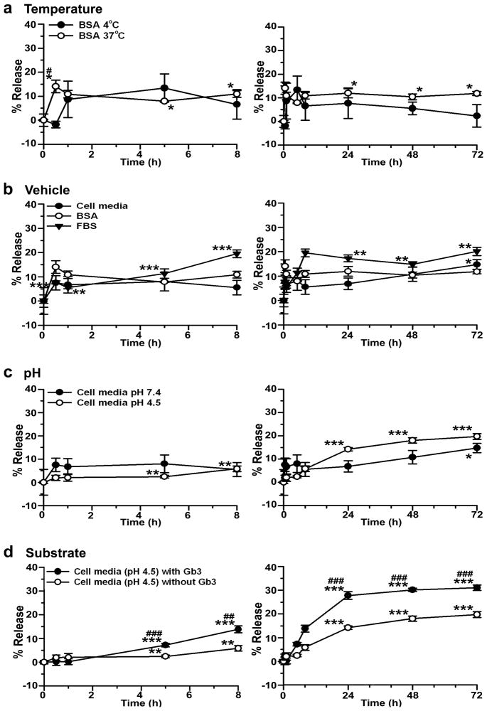
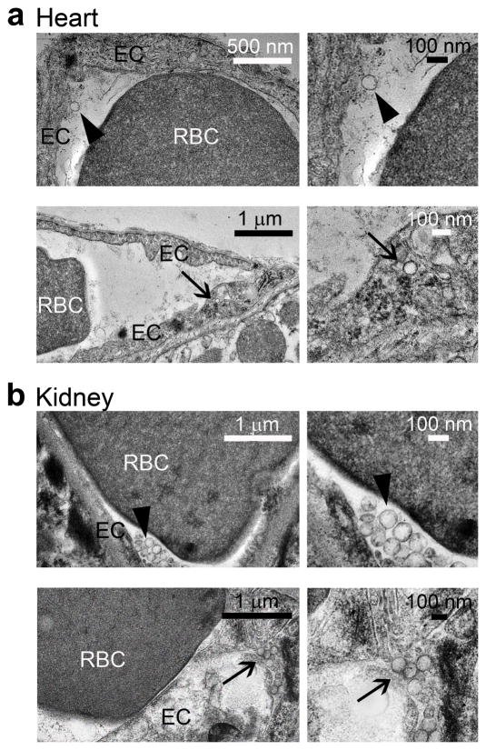
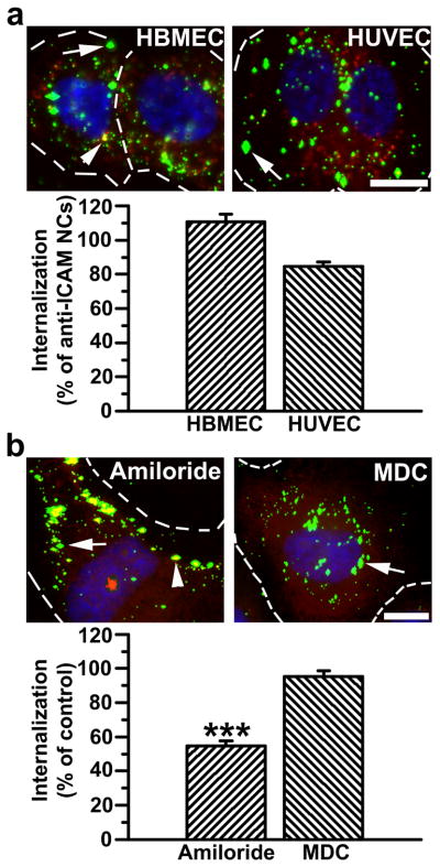
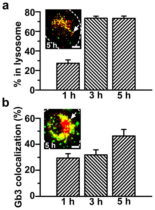
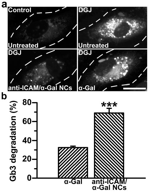
Fabry disease, due to the deficiency of α-galactosidase A (α-Gal), causes lysosomal accumulation of globotriaosylceramide (Gb3) in multiple tissues and prominently in the vascular endothelium. Although enzyme replacement therapy (ERT) by injection of recombinant α-Gal improves the disease outcome, the effects on the vasculopathy associated with life-threatening cerebrovascular, cardiac and renal complications are still limited. We designed a strategy to enhance the delivery of α-Gal to organs and endothelial cells (ECs). We targeted α-Gal to intercellular adhesion molecule 1 (ICAM-1), a protein expressed on ECs throughout the vasculature, by loading this enzyme on nanocarriers coated with anti-ICAM (anti-ICAM/α-Gal NCs). In vitro radioisotope tracing showed efficient loading of α-Gal on anti-ICAM NCs, stability of this formulation under storage and in model physiological fluids, and enzyme release in response to lysosome environmental conditions. In mice, the delivery of (125)I-α-Gal was markedly enhanced by anti-ICAM/(125)I-α-Gal NCs in brain, kidney, heart, liver, lung, and spleen, and transmission electron microscopy showed anti-ICAM/α-Gal NCs attached to and internalized into the vascular endothelium. Fluorescence microscopy proved targeting, endocytosis and lysosomal transport of anti-ICAM/α-Gal NCs in macro- and micro-vascular ECs and a marked enhancement of Gb3 degradation. Therefore, this ICAM-1-targeting strategy may help improve the efficacy of therapeutic enzymes for Fabry disease.


Figures

Figure 1
Figure 1. Release of α-Gal from anti-ICAM nanocarriers
125I-α-Gal released from anti-ICAM/125I-α-Gal NCs was separated from the particle-bound fraction by centrifugation after incubation for varying periods of time in (a) 1% BSA in PBS buffer at 4°C or 37°C; (b) 1% BSA in PBS, HUVEC medium, or FBS at 37°C and pH 7.4; (c) HUVEC medium at 37°C and either pH 7.4 or pH 4.5; and (d) HUVEC medium at 37°C and pH 4.5 in the absence or presence of NBD-Gb3. Data are mean±SEM (n≥3). * compares each time point to time 0 within the same condition. # compares (for each time point): (a) 4°C to 37°C, (b) HUVEC medium and FBS to BSA, (c) pH 4.5 to pH 7.4, and (d) Gb3 to non-Gb3. * and # are p≤0.05; ** and ## are p≤0.01; *** and ### are p≤0.001, by Student’s t-test. Statistical significance for time 0–8 h are shown only in the left panel and time 24–72 h only in the right panel.
Figure 2
Figure 2. Visualization of anti-ICAM/α-Gal nanocarriers in mice
Transmission electron micrographs showing the presence of anti-ICAM/α-Gal NCs in (a) heart and (b) kidney, both attached to the endothelial surface (arrowheads, upper panels) and internalized within cells (arrows, bottom panels) by 30 min after injection.
Figure 3
Figure 3
Efficient targeting and internalization of anti-ICAM/α-Gal nanocarriers in micro- and macro-vascular endothelial cells. (a) Fluorescence microscopy images and quantification of TNFα-activated HBMECs and HUVECs incubated with FITC-labeled anti-ICAM/α-Gal NCs at 37°C for 30 min, washed and incubated in cell medium for 30 min. Cells were fixed and surface-bound NCs and nuclei were stained with Texas-Red-labeled anti-mouse IgG and DAPI, respectively. Internalization was compared to that of anti-ICAM NCs, shown in Figure S2. (b) Uptake of anti-ICAM/α-Gal NCs was also tested in the presence of amiloride or MDC, which inhibit CAM- vs clathrin-mediated endocytosis, respectively. In both (a) and (b), single-labeled green NCs are internalized (arrow) vs double-labeled (green+red) yellow NCs, which are located in the cell surface (arrowhead). Dashed lines mark the cell border, determined by phase-contrast. Scale bar, 10 μm. Data are mean±SEM (n≥55 cells, duplicated). *** is p≤0.001, by Student’s t-test.
Figure 4
Figure 4. Internalized anti-ICAM/α-Gal nanocarriers traffic to lysosomes and colocalize with Gb3
(a) TNFα-activated HUVECs were incubated at 37°C for 1 h with Texas-Red dextran to label lysosomes, followed by incubation with FITC-labeled anti-ICAM/α-Gal NCs for 1 hour at 37°C. Cells were washed and fixed, or washed and incubated with cell medium for 2 or 4 additional hours (total incubation time: 1, 3, and 5 h) and then fixed. (b) Cells were incubated for 16 h with DGJ and NBD-Gb3 to visualize accumulation of this green-fluorescent substrate analog in intracellular compartments (red pseudocolor) and then incubated with non-fluorescent anti-ICAM/α-Gal NCs as in a. Cells were then washed, fixed, and permeabilized, and NCs were stained with Texas-Red-labeled goat anti-mouse IgG (green pseudocolor). Samples were analyzed by fluorescence microscopy to determine the percentage of (pseudo) green-labeled NCs which colocalized within (pseudo) red-labeled lysosomes or Gb3-positive compartments (yellow, arrows). Dashed lines mark the cell borders, determined by phase-contrast. Scale bar, 10 μm. Data are mean±SEM (n≥23 cells, duplicated).
Figure 5
Figure 5. Anti-ICAM/α-Gal nanocarriers attenuate Gb3 accumulation in an endothelial cell model of Fabry disease
(a) TNFα-activated HUVECs were incubated at 37°C for 16 h with fluorescent NBD-Gb3 and control cell medium or medium containing DGJ to inhibit endogenous αGal. Cells were then washed and left untreated or treated with α-Gal or non-fluorescent anti-ICAM/α-Gal NCs for 5 h, all in the presence of chloroquine to selectively permit activity of exogenous neutral α-Gal, and not endogenous acidic α-Gal. Cells were fixed and analyzed by fluorescence microscopy. Dashed lines mark cell borders, determined by phase-contrast. Scale bar, 10 μm. (b) Intracellular accumulation of NBD-Gb3 was quantified from micrographs as described in Materials and Methods. Data are mean±SEM (n≥72 cells). *** is p≤0.001, by Student’s t-test.

References

    1. Desnick RJ, Ioannou YA, Eng CM. In: XVI Chapter, Lysosomal Disorders. The Metabolic and Molecular Bases of Inherited Disease. 8. Scriver C, Beaudet A, Sly W, Valle D, Childs B, Kinzler K, Vogelstein B, editors. McGraw-Hill; 2001.
    1. Park JL, Whitesall SE, D’Alecy LG, Shu L, Shayman JA. Vascular dysfunction in the alpha-galactosidase A-knockout mouse is an endothelial cell-, plasma membrane-based defect. Clin Exp Pharmacol Physiol. 2008;35(10):1156–1163. - PMC - PubMed
    1. Shu L, Shayman JA. Caveolin-associated accumulation of globotriaosylceramide in the vascular endothelium of alpha-galactosidase A null mice. J Biol Chem. 2007;282(29):20960–20967. - PubMed
    1. Lee K, Jin X, Zhang K, Copertino L, Andrews L, Baker-Malcolm J, Geagan L, Qiu H, Seiger K, Barngrover D, McPherson JM, Edmunds T. A biochemical and pharmacological comparison of enzyme replacement therapies for the glycolipid storage disorder Fabry disease. Glycobiology. 2003;13(4):305–313. - PubMed
    1. Kornfeld S, Reitman ML, Varki A, Goldberg D, Gabel CA. Steps in the phosphorylation of the high mannose oligosaccharides of lysosomal enzymes. Ciba Found Symp. 1982;(92):138–156. - PubMed

Publication types

MeSH terms