. 2010 Dec;8(12):1591-600.
doi: 10.1158/1541-7786.MCR-10-0209. Epub 2010 Oct 27.

RGS-GAIP-interacting protein controls breast cancer progression

Affiliations

RGS-GAIP-interacting protein controls breast cancer progression

Ling Wang et al. Mol Cancer Res. 2010 Dec.

Abstract

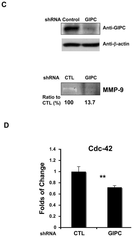
Although the importance of RGS-GAIP-interacting protein (GIPC) in the biology of malignant cells is well known, the molecular mechanism of GIPC in the inhibition of tumor progression has not been identified. This study focused on elucidating the molecular role of GIPC in breast cancer progression. By using a human breast tumor specimen, an in vivo mouse model, and breast cancer cell lines, we showed for the first time that GIPC is involved in breast cancer progression through regulation of breast cancer cell proliferation, survival, and invasion. Furthermore, we found that the Akt/Mdm2/p53 axis, insulin-like growth factor-1 receptor, matrix metalloproteinase-9, and Cdc42 were downstream of GIPC signaling in breast cancer cells. Moreover, we showed that wild-type p53 reduced GIPC-induced breast cancer cell survival, whereas mutant p53 inhibited GIPC-induced cell invasion. Finally, we demonstrated that an N-myristoylated GIPC peptide (CR1023, N-myristoyl-PSQSSSEA) capable of blocking the PDZ domain of GIPC successfully inhibited MDA-MB-231 cell proliferation, survival, and further in vivo tumor growth. Taken together, these findings demonstrate the importance of GIPC in breast tumor progression, which has a potentially significant impact on the development of therapies against many common cancers expressing GIPC, including breast and renal cancer.


Figures

Figure 1
Figure 1. Role of GIPC in breast cancer progression
A. The expression of GIPC in human breast cancer specimens via immunohistochemical staining. Staining of GIPC in normal breast tissue (NBT) (a); Staining of GIPC in a moderately differentiated infiltrating ductal carcinoma (IDC) (b); Staining of GIPC in an undifferentiated IDC (c). The insets show a magnification of GIPC “luminal” staining pattern in human breast tumor specimens. B. The absence of GIPC decreased tumor volume in vivo. GIPC knockdown was confirmed in the cells prior to injection (upper panel).
Figure 2
Figure 2. Tumorigenic properties of GIPC in breast cancer development
A. Western blots showing native expression levels of GIPC in the breast cancer cell lines MDA-MB-231 and MCF-7. Human umbilical vein endothelial cells (HUVECs) were used as a positive control. B. GIPC mediates breast cancer cell viability and proliferation as assessed through an MTS assay. *p<0.01, in a student’s t test compared with control (CTL). Western blot analysis confirmed GIPC expression after siRNA knockdown (upper panel). C. MCF-7 cells with downregulated GIPC expression showed an increase in apoptosis. No significant difference was observed in MDA-MB-231 cells. *p<0.05, **p<0.01 in a student’s t test. D. NRP-1 and GIPC knockdown reduced migration and invasion in MDA-MB-231 cells. *p<0.01, **p<0.001 in a student’s t test compared with control (CTL).
Figure 3
Figure 3. The effect of GIPC knockdown in MDA-MB-231 and MCF-7 cells
A number of molecules were analyzed after GIPC knockdown: A. Phosphorylated Akt, phosphorylated Mdm2, and p53; B. IGF-1Rβ and phospho-IGF-1Rβ in Western blot. β-actin was used as the loading control. The fold difference of p-IGF-1Rβ is normalized to β-actin expression; C. MMP-9 activity was assessed by gelatin zymography; D. Cdc-42 was analyzed.
Figure 4
Figure 4. p53 status determines the role of GIPC in MDA-MB-231
A. Wild-type p53 (wt-p53) mediates MCF-7 cell survival through GIPC. *p<0.05 and **p<0.01 compared with CTL siRNA group; #p<0.05 and ##p<0.01 compared with GIPC siRNA group as determined by the student’s t test; B. Mutant p53 (mut-p53) is involved in GIPC invasion signaling. In cells lacking both GIPC and mutant p53, invasion levels recovered. *p<0.05 compared with the control siRNA group; #p<0.01 compared with the GIPC siRNA group as determined by the student’s t test; C. MMP-9 activity was assessed in supernatants from GIPC depleted MDA-MB-231 cells treated with p53 siRNA. Activity was quantified as a ratio to the CTL sample. Expression levels of p53 and GIPC are shown in the upper panel.
Figure 5
Figure 5. Myristylated GIPC peptide CR1023 treatment regulates IGF-1R expression, inhibits proliferation and cell viability
A. As determined by Western blotting, the expression level of IGF-1R decreased with CR1023 treatment; B. With a [3H]-thymidine incorporation assay, a dose-dependent inhibition of cell proliferation was observed in samples treated with CR1023. No inhibition was observed with control CR2055 peptide treatment; C. Apoptosis assay of MDA-MB-231-WT cells treated with DMSO, CR1023 and CR2055 in a dose-dependent manner for 48 hours. A dose-dependent increase in apoptosis was observed after treatment with CR1023. No inhibition was observed with either the control peptide CR2055 or DMSO.
Figure 6
Figure 6. In vivo effect of intratumoral injection of peptide on tumor weight, volume, and IGF-1R expression
A. Effect of administration of CR1023 peptide on tumor growth over time; B and C. Tumor weight and tumor volumes decreased after CR1023 treatment compared with DMSO treatment alone; D. IGF-1R expression is decreased in harvested tumors after CR1023 treatment (T) compared with tumors treated with DMSO alone (U).

Similar articles

Cited by

References

    1. De Vries L, Lou XJ, Zhao G, Zheng B, Farquhar MG. GIPC, a PDZ domain containing protein, interacts specifically with the C terminus of RGS-GAIP. Proceedings of the National Academy of Sciences of the United States of America. 1998;95:12340–5. - PMC - PubMed
    1. Kedlaya RH, Bhat KM, Mitchell J, Darnell SJ, Setaluri V. TRP1 interacting PDZ-domain protein GIPC forms oligomers and is localized to intracellular vesicles in human melanocytes. Arch Biochem Biophys. 2006;454:160–9. - PMC - PubMed
    1. Kirikoshi H, Katoh M. Expression of human GIPC1 in normal tissues, cancer cell lines, and primary tumors. Int J Mol Med. 2002;9:509–13. - PubMed
    1. Muders MH, Dutta SK, Wang L, Lau JS, Bhattacharya R, Smyrk TC, et al. Expression and regulatory role of GAIP-interacting protein GIPC in pancreatic adenocarcinoma. Cancer Research. 2006;66:10264–8. - PubMed
    1. Awan A, Lucic MR, Shaw DM, Sheppard F, Westwater C, Lyons SA, et al. 5T4 interacts with TIP-2/GIPC, a PDZ protein, with implications for metastasis. Biochem Biophys Res Commun. 2002;290:1030–6. - PubMed

Publication types

MeSH terms

Substances