. 2011 Mar;39(5):1680-91.
doi: 10.1093/nar/gkq900. Epub 2010 Nov 2.

The effects of histone H4 tail acetylations on cation-induced chromatin folding and self-association

Affiliations

The effects of histone H4 tail acetylations on cation-induced chromatin folding and self-association

Abdollah Allahverdi et al. Nucleic Acids Res. 2011 Mar.

Abstract

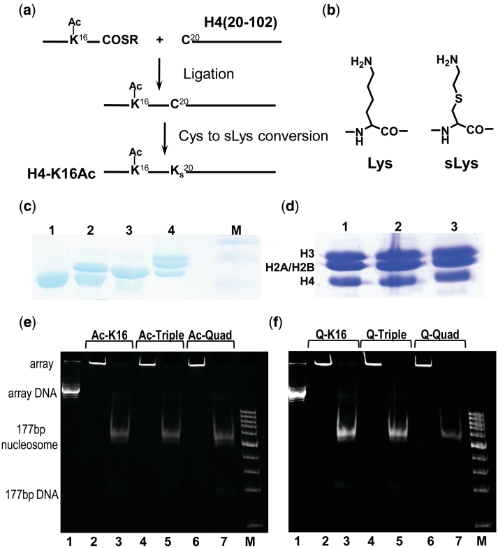
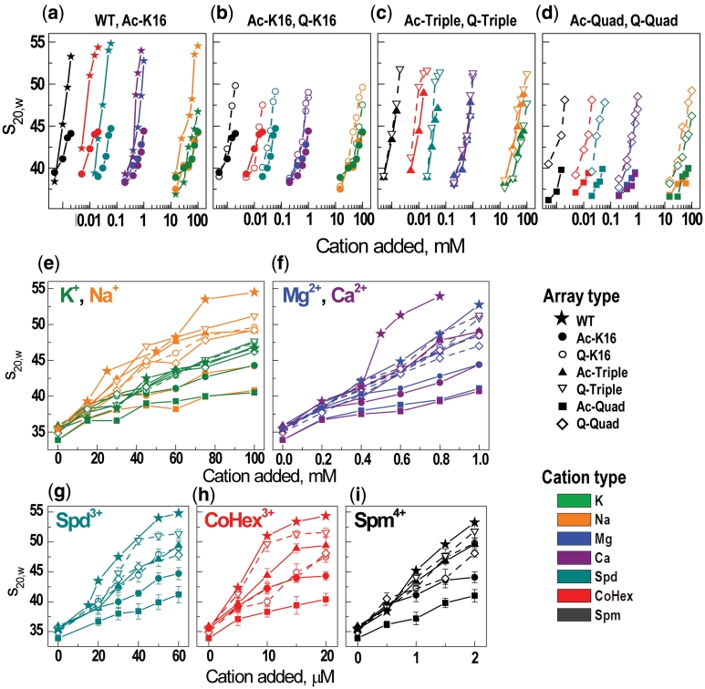
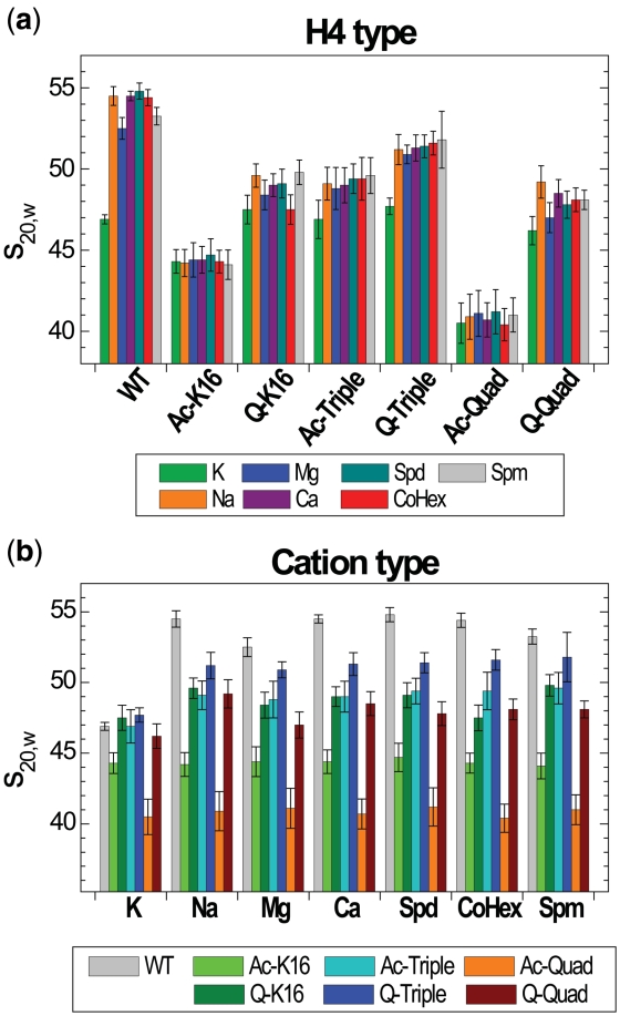
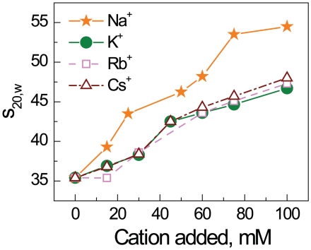
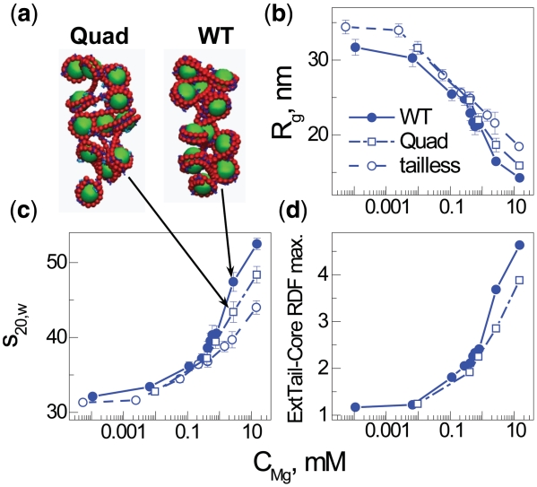
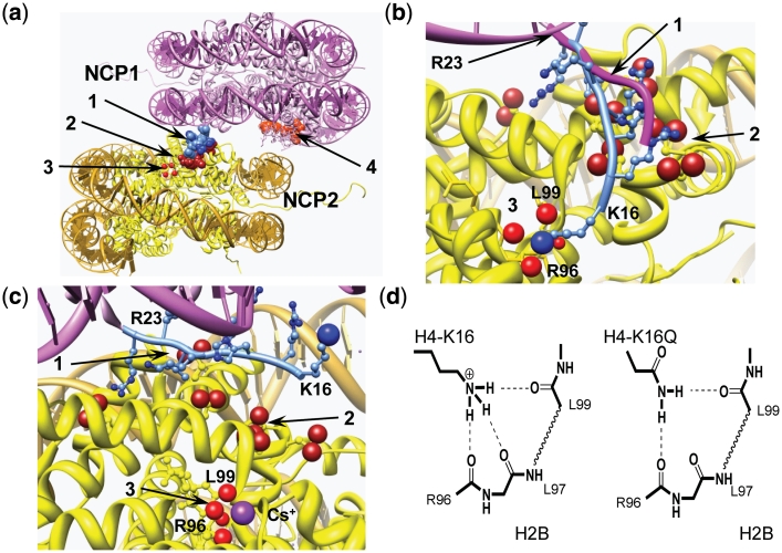
Understanding the molecular mechanisms behind regulation of chromatin folding through covalent modifications of the histone N-terminal tails is hampered by a lack of accessible chromatin containing precisely modified histones. We study the internal folding and intermolecular self-association of a chromatin system consisting of saturated 12-mer nucleosome arrays containing various combinations of completely acetylated lysines at positions 5, 8, 12 and 16 of histone H4, induced by the cations Na(+), K(+), Mg(2+), Ca(2+), cobalt-hexammine(3+), spermidine(3+) and spermine(4+). Histones were prepared using a novel semi-synthetic approach with native chemical ligation. Acetylation of H4-K16, but not its glutamine mutation, drastically reduces cation-induced folding of the array. Neither acetylations nor mutations of all the sites K5, K8 and K12 can induce a similar degree of array unfolding. The ubiquitous K(+), (as well as Rb(+) and Cs(+)) showed an unfolding effect on unmodified arrays almost similar to that of H4-K16 acetylation. We propose that K(+) (and Rb(+)/Cs(+)) binding to a site on the H2B histone (R96-L99) disrupts H4K16 ε-amino group binding to this specific site, thereby deranging H4 tail-mediated nucleosome-nucleosome stacking and that a similar mechanism operates in the case of H4-K16 acetylation. Inter-array self-association follows electrostatic behavior and is largely insensitive to the position or nature of the H4 tail charge modification.


Figures

Figure 1.
Figure 1.
(a and b) Scheme for the semi-synthesis preparation of histones with covalently modified histone N-terminal tails by Native Chemical Ligation (NCL) in combination with S-alkylation. Amino acids 1–19 of the histone H4 with lysines acetylated at selected positions were manually synthesized by the Boc-based SPPS method and ligated to the recombinantly prepared variant of the H4 histone [amino acids 20–102 with K20C mutation, H4(20-102)K20C]. (a) Preparation of H4K16-Ac. First step: Ligation to form an Arg19-Cys20 junction. Second step: Conversion of Cys20 to sLys20 by treatment with bromoethylamine. (b) Comparison of Lys and sLys. (c) 18% SDS–PAGE illustrating production of the HO with H4-K16Ac histone: lane 1—globular H4(20-102)K20C (before ligation); lane 2—mixture of H4(20-102)K20C and ligation product (H4(K16Ac,K20C); lane 3—purified H4 K16Ac; 4—HO with H4 K16Ac; M—marker. (d) 18% SDS–PAGE of the HOs with acetylated forms of the histone H4. Lane 1, HO with tetra-acetylated H4 K5,8,12,16Ac; lane 2, HO with tri-acetylated H4 K5,8,12 Ac; lane 3, HO with H4 K16Ac. (e and f) Characterization of purity and saturation of the 12-177-601 nucleosome arrays containing (e) acetylated and (f) K→Q mutated histone H4. Array identity is indicated on the top of the lanes. In (e and f), lanes 2,4,6 are for the ScaI untreated array; lanes 3, 5 and 7 for the array after ScaI digestion; lanes 1 is 12-177-601 DNA; M is DNA marker from 100 to 1500 bp, step 100 bp.
Figure 2.
Figure 2.
Summary of the AUC results: (a–d) Pair-wise (indicated at the top of each graph) comparison of the cation concentration dependencies of the s20,w values for the array containing recombinant (WT), selectively acetylated, or K→Q mutated H4 histone. Graphs a–d are built in the same scale with origin of y-axis placed at s20,w = 35.2S (an average of the s20,w values measured for all arrays in the TEK buffer with no added cation). (e–i) AUC titration curves with a linear scale of cation concentration and grouped according to the cation charge and nature (indicated at the top of each graph). For all graphs in the figure, symbols vary for the different variants of the H4 histone; color coding is used for different cations.
Figure 3.
Figure 3.
Maximal s20,w values measured upon increase cation concentration in the array solution grouped (a) by the type of H4 histone or (b) by the cation. Color coding for the H4 or cation type is indicated at the bottom of respective graph.
Figure 4.
Figure 4.
AUC sedimentation velocity results for the recombinant WT 12-177-601 nucleosome array in the presence of various concentrations of alkali metal chlorides, NaCl, KCl, RbCl and CsCl.
Figure 5.
Figure 5.
Summary of coarse-grained computer simulations of the 12–177 nucleosome array with added Mg2+. Data obtained for three different models of the array mimicking the WT, Quad or tailless array are shown as indicated in the (b) graph legend. Data calculated for an array with a single charge quenched in the histone H4 tail mimicking a single modification are not displayed since they are very close to the data of the WT array. Representative snapshots in (a) show folding of the Quad (left) and WT (right) arrays at bulk Mg2+ concentration 2.7 mM, demonstrating the moderate effect on folding caused by quenching the four charges K5,8,12,16 of the histone H4 tail. Dependencies of radius of gyration, Rg, (b); sedimentation velocity coefficient, s20,w, (c); and intensity of the maximum in the external tail—central core particle RDF (d) on bulk concentration of Mg2+. The external tail-core RDF signals the propensity of tail bridging to neighboring nucleosomes. Bulk concentration of Mg2+ is defined as concentration of Mg2+ ions averaged over the regions of simulation cell with low electric filed from the nucleosome array (32).
Figure 6.
Figure 6.
Summary of the PA results. (a) Precipitation curves showing absorbance A260 in supernatant of the array solution (10 mM Tris–HCl, pH 7.5; initial array concentration CP = 151 µM of DNA) versus concentration of the added cations Mg2+, Ca2+, Spd3+, CoHex3+ and Spm4+ (indicated at the top of each graph; addition of K+ or Na+ does not result in precipitation of the array <50%, data not shown). Points are measured values (normalized relative to the A260 absorbance in the array solution without added cation); curves are sigmoidal fitting of the experimental data. (b) EC50 values of the 12-177-601 arrays with the WT, acetylated and K→Q mutated histone H4 obtained for various cations (indicated in the graphs).
Figure 7.
Figure 7.
Structural analysis of NCP–NCP stacking. (a) NCP–NCP contact in the crystal 1AOI (1) with important domains involved in formation of the contact shown in space filling: 1 light blue, H4 K16-R23 of the NCP1; 2 dark red, acidic islet of the H2A histone (amino acids E56, E61, E64, D90, E91, E92) in the NCP2 interacting with H4 K16-R23 domain of the NCP1; 3 four red spheres, carbonyl atoms of the main chain peptide H2B R96-L99 of the NCP2; 4 light red, acidic islet of the H2A histone of the NCP1 needed to be screened in stacking contact. (b) Detailed presentation of the H4 K16-R23 binding to the neighboring NCP2 with hypothetical relocation of the H4-K16 from the acidic H2A patch to the H2B R96-L99-binding site. To accommodate the H4-K16 ε-amino group coordination with the H2B R96-L99 carbonyl groups, the NCP1 was moved 2 Å towards the H2B site and the structure of the H4 K16-R23 fragment (shown in light blue with H4-K16 ε-amino group as a blue sphere) was engineered by manipulating torsion angles. The original location of the H4 K16-R23 chain in the 1AOI crystal is indicated by magenta tracing of the peptide backbone. (c) The same region of the NCP–NCP contact as in (b) but built using the crystal structure of the NCP saturated with Cs+ [3MGS (58)]. The Cs+ ion coordinated with R96, L97, L99 carbonyl oxygen atoms is seen at the bottom of the cartoon. In (b) and (c), domains are numbered as in (a) with charged oxygen atoms of the carboxylate group in the acidic islet of the H2A histone highlighted as red spheres. (d) Proposed hydrogen bonding of the H4-K16 binding to the crystallographically determined Rb+/Cs+-binding pocket in (c). To the left is shown the proposed native K16 ε-amine hydrogen bonding and the right demonstrates alternative hydrogen bonding upon K→Q mutation of H4-K16, which maintains this structural element. Acetylation is suggested to completely disrupt this binding due to steric constraints and reduced H-bonding capacity. Chimera visualization and molecule editing package (63) was used to build the structures in a–c.

References

    1. Luger K, Mader AW, Richmond RK, Sargent DF, Richmond TJ. Crystal structure of the nucleosome core particle at 2.8 Å resolution. Nature. 1997;389:251–260. - PubMed
    1. Harp JM, Hanson BL, Timm DE, Bunick GJ. Asymmetries in the nucleosome core particle at 2.5 Å resolution. Acta Cryst. Sect. D. 2000;56:1513–1534. - PubMed
    1. Davey CA, Sargent DF, Luger K, Maeder AW, Richmond TJ. Solvent mediated interactions in the structure of nucleosome core particle at 1.9 Å resolution. J. Mol. Biol. 2002;319:1097–1113. - PubMed
    1. Dorigo B, Schalch T, Kulangara A, Duda S, Schroeder RR, Richmond TJ. Nucleosome arrays reveal the two-start organization of the chromatin fiber. Science. 2004;306:1571–1573. - PubMed
    1. Robinson PJJ, Fairall L, Huynh VAT, Rhodes D. EM measurements define the dimensions of the ‘30-nm’ chromatin fiber: evidence for a compact, interdigitated structure. Proc. Natl Acad. Sci. USA. 2006;103:6506–6511. - PMC - PubMed

Publication types