. 2011 Jan;85(1):334-47.
doi: 10.1128/JVI.01278-09. Epub 2010 Nov 3.

The Tyro3 receptor kinase Axl enhances macropinocytosis of Zaire ebolavirus

Affiliations

The Tyro3 receptor kinase Axl enhances macropinocytosis of Zaire ebolavirus

Catherine L Hunt et al. J Virol. 2011 Jan.

Abstract

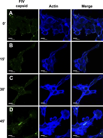
Axl, a plasma membrane-associated Tyro3/Axl/Mer (TAM) family member, is necessary for optimal Zaire ebolavirus (ZEBOV) glycoprotein (GP)-dependent entry into some permissive cells but not others. To date, the role of Axl in virion entry is unknown. The focus of this study was to characterize entry pathways that are used for ZEBOV uptake in cells that require Axl for optimal transduction and to define the role of Axl in this process. Through the use of biochemical inhibitors, interfering RNA (RNAi), and dominant negative constructs, we demonstrate that ZEBOV-GP-dependent entry into these cells occurs through multiple uptake pathways, including both clathrin-dependent and caveola/lipid raft-mediated endocytosis. Other dynamin-dependent and -independent pathways such as macropinocytosis that mediate high-molecular-weight dextran uptake also stimulated ZEBOV-GP entry into these cells, and inhibitors that are known to block macropinocytosis inhibited both dextran uptake and ZEBOV infection. These findings provided strong evidence for the importance of this pathway in filovirus entry. Reduction of Axl expression by RNAi treatment resulted in decreased ZEBOV entry via macropinocytosis but had no effect on the clathrin-dependent or caveola/lipid raft-mediated endocytic mechanisms. Our findings demonstrate for the first time that Axl enhances macropinocytosis, thereby increasing productive ZEBOV entry.


Figures

FIG. 1.
FIG. 1.
Axl is necessary for efficient infectious Zaire ebolavirus infection and FIV-ZEBOV transduction into Axl-dependent cells. (A and C) Effect of Axl RNAi on ZEBOV infection or transduction. SNB19 cells were transfected with 200 pmol of a nonspecific luciferase siRNA control (A), 200 pmol of a nonspecific siRNA control (Block-It) (C), or 200 pmol of a human Axl-specific siRNA (A and C). At 48 h following RNAi transfection, cells were infected with ZEBOV (A) (MOI, 0.25) for 24 h or transduced with FIV pseudovirions for 48 h (C) (MOI, 0.005). At 24 h following infection, cells were fixed and analyzed by microscopy for GFP positivity relative to the number of cells for each condition (A), or at 48 h following transduction, cells were fixed and stained for β-Gal activity (C). (B and D) Knockdown of Axl by RNAi. At 48 h after RNAi transfection, a portion of the transfected cells were lysed, proteins were separated using SDS-PAGE, and Axl was detected on the nitrocellulose membrane with primary antiserum (1:4,000) overnight at 4°C. The signal was detected by incubation with secondary horseradish peroxidase-conjugated antiserum (1:20,000) for 1 h at room temperature followed by visualization by chemiluminescence. Actin was detected by incubation with horseradish peroxidase-conjugated anti-actin antibody. The immunoblots shown are representative of three experiments performed independently. (E) Ability of Axl antiserum to block transduction of FIV-ZEBOV or FIV-MARV. A 1:50 dilution of goat anti-human Axl antiserum or normal goat serum was incubated with SNB19 cells for 1 h at 4°C. VSVΔG pseudovirions (MOI, 0.05) bearing full-length ZEBOV-GP or MARV-GP were applied in the presence of antiserum and incubated for an additional 23 h. Cells were lifted and analyzed by flow cytometry for GFP positivity, indicating viral transduction. Data represent the averages and standard errors of three experiments performed in triplicate. **, P < 0.001.
FIG. 2.
FIG. 2.
Clathrin- and caveola/lipid raft-mediated endocytic pathways facilitate FIV-ZEBOV transduction of Axl-dependent cells. (A) Ability of CPZ to inhibit FIV-ZEBOVΔO transduction of SNB19 cells. Cells were pretreated with the indicated amounts of CPZ for 1 h. Treated SNB19 cells were incubated with FIV-ZEBOVΔO (MOI, 0.005), FIV-VSV-G (MOI, 0.005), Cy5-Tfr (20 μg/ml), or Cy5-CTb (10 μg/ml) in the continued presence of CPZ. Tfr- or CTb-treated SNB19 cells were washed after 1 h and analyzed by flow cytometry for uptake of Tfr or CTb. The medium on cells transduced with FIV-ZEBOVΔO or FIV-VSV-G was refreshed after 6 h with medium not containing CPZ, and transduced cells were fixed and stained for β-Gal activity 48 h following transduction. (B) Ability of DNEps15 to inhibit FIV-ZEBOVΔO transduction. SNB19 cells were transfected with plasmid DNA expressing eGFP, wild-type Eps15-GFP (wtEps15), or dominant negative Eps-GFP (DNEps15). The transfection efficiency of SNB19 cells was evaluated at 24 h by analyzing GFP expression in the cells by flow cytometry and was found to be between 50 and 60%. Transfected cells were transduced with FIV-ZEBOVΔO (MOI, 0.005) or FIV-VSV-G (MOI, 0.005) at 24 h following transfection. After an additional 48 h, cells were fixed and stained for β-Gal activity. Alternatively, transfected cells were incubated for 1 h with Cy5-labeled Tfr or CTb and washed and cells were analyzed by flow cytometry for uptake of Tfr and CTb. (C) Ability of CPZ to inhibit FIV-ZEBOV transduction in Hffs. Studies were performed as described for panel A. (D) Ability of FIL to inhibit FIV-ZEBOVΔO transduction into SNB19 cells. Cells were pretreated with the indicated amounts of FIL for 1 h in serum-free medium. The FIL was removed, and cells were incubated with FIV-ZEBOVΔO (MOI, 0.005), FIV-VSV-G (MOI, 0.005), or Cy5-labeled Tfr or CTb in serum-free medium. Tfr- or CTb-treated SNB19 cells were washed after 1 h and analyzed by flow cytometry for uptake of Tfr or CTb. The medium on cells transduced with FIV-ZEBOVΔO or FIV-VSV-G was refreshed after 6 h with medium containing serum without FIL, and transduced cells were fixed and stained for β-Gal activity 48 h following transduction. (E) Knockdown of Cav1/2 inhibits FIV-ZEBOVΔO transduction in SNB19 cells. Cells were transfected with 200 pmol of a nonspecific siRNA control (Block-It) or 200 pmol of a mixture of human Cav1 and Cav 2 siRNAs. At 48 h after transfection, cells were incubated with FIV-ZEBOVΔO, FIV-VSV-G pseudovirions (MOI, 0.005), or Cy5-conjugated Tfr or CTb. After 1 h, the SNB19 cells were analyzed by flow cytometry for uptake of Tfr and CTb. At 48 h following transduction, transduced SNB19 cells were fixed and stained for β-Gal activity. (F) Knockdown of Cav1/2 in SNB19 cells by RNAi. At 48 h after transfection, a portion of the cells were lysed and proteins were separated using SDS-PAGE. Cav1 and Cav2 were detected on the nitrocellulose membrane with primary antibodies (1:1,000 and 1:250, respectively) incubated overnight at 4°C followed by incubation with horseradish peroxidase-conjugated secondary antiserum (1:20,000) for 1 h at room temperature. The blot was visualized by chemiluminescence. Actin was detected by incubation with horseradish peroxidase-conjugated anti-actin antibody (1:10,000). (G) Knockdown of Cav1/2 inhibits FIV-ZEBOVΔO transduction in Hffs. Studies were performed as described for panel E. Cell viability in the presence of the various treatments is shown as a dotted line in panels A to G. Data represent the averages and standard errors of three experiments performed in triplicate. *, P < 0.05; **, P < 0.001. EtOH, ethanol.
FIG. 3.
FIG. 3.
Dynamin is necessary for efficient ZEBOV-GP pseudovirion entry into Axl-dependent cells. (A to C) Ability of DN dynamin to inhibit FIV-ZEBOVΔO transduction. SNB19 cells (A), Hffs (B), and HuVECs (C) were transduced with adenoviral vectors bearing GFP (Ad-GFP) or a dominant negative form of dynamin 2 (Ad-DN-dyn2) at an MOI of 30 for SNB19 cells and an MOI of 90 for Hffs and HuVECs, ensuring greater than 90% adenovirus transduction. Eighteen hours following adenoviral transduction, SNB19 (A) and Hff (B) cells were incubated with FITC-labeled dextran (0.5-mg/ml final concentration) for 1 h or transduced with FIV pseudovirions (MOI, 0.005; all cell populations) for 48 h. SNB19 and Hff cells were analyzed by flow cytometry for uptake of dextran after 1 h. Transduced cells were fixed and stained for β-Gal activity after 48 h. (D) Ability of Dynasore to inhibit FIV-ZEBOVΔO transduction. SNB19 cells were treated for 1 h with the indicated amounts of Dynasore. Treated cells were incubated with FITC-conjugated dextran or Cy5-conjugated Tfr or CTb for 1 h and analyzed by flow cytometry or incubated with FIV pseudovirions (MOI, 0.005) for an additional 6 h in the presence of the drug. The medium on cells transduced with FIV-ZEBOVΔO or FIV-VSV-G was refreshed after 6 h with medium not containing drug, and transduced cells were fixed and stained for β-Gal activity 48 h following transduction. Cell viability in the presence of the treatments is shown as a dashed line in all panels. Data represent the averages and standard errors of three experiments performed in triplicate. *, P < 0.05; **, P < 0.001.
FIG. 4.
FIG. 4.
Inhibitors of macropinocytosis decrease ZEBOV infection in Axl-dependent cells. (A) Ability of macropinocytosis inhibitors to inhibit dextran but not Tfr and CTb uptake. SNB19 cells were treated for 1 h with the indicated amounts of macropinocytosis inhibitors. After 1 h, Cy5-Tfr, Cy5-CTb, or FITC-dextran was added to the cells in the presence of drug for an additional 1 h. The cells were then analyzed by flow cytometry for uptake of the labeled conjugates. (B to D) Ability of macropinocytosis inhibitors to inhibit FIV-ZEBOV transduction. SNB19 cells were incubated for 1 h with the indicated concentrations of macropinocytosis inhibitors. After 1 h, the cells were transduced with FIV pseudovirions (MOI, 0.005) in the presence of the drug for an additional 6 h. Medium was refreshed without drug, and cells were incubated for an additional 48 h. Cells were fixed and stained for β-Gal activity. (E) Ability of macropinocytosis inhibitors to inhibit ZEBOV virus-like particle (VLP) entry into SNB19 cells. SNB19 cells were treated for 1 h with the indicated amounts of macropinocytosis inhibitors. After 1 h, ZEBOV VLPs were added to cells in the presence of drug for an additional 6 h. Unbound VLPs were removed, and cells were lysed and analyzed for luciferase activity. (F and G) Ability of macropinocytosis inhibitors to decrease ZEBOV infection. SNB19 cells were incubated with the indicated concentrations of drug for 1 h. Infectious ZEBOV (MOI, 0.25) was added in the presence of the drug for an additional 17 h. At 17 h following ZEBOV infection, the medium was changed on the cells, and 6 h after that, cells were fixed and assessed by microscopy for GFP positivity relative to the number of cells present for each condition. (H) Indicated amounts of macropinocytosis inhibitors were incubated with SNB19 cells for 1 h. After 1 h, SNB19 cells were transduced with VSVΔG full-length ZEBOV or VSVΔG-MARV-GP (MOI, 0.05). Following 6 h of incubation with virus in medium containing drug, the medium was removed and replaced with medium without drug. VSV-transduced cells were assessed for GFP expression by flow cytometry at 23 h following transduction. Data represent the averages and standard errors of three experiments performed in triplicate. *, P < 0.05; **, P < 0.001.
FIG. 5.
FIG. 5.
Actin polymerization increases during FIV-ZEBOVΔO pseudovirion transduction of Axl-dependent SNB19 cells. (A to D) Confocal microscopy of FIV capsid (green) and F-actin (blue) during FIV-ZEBOVΔO pseudovirion transduction (MOI, 250) for 1 h at 4°C (A) or 1 h at 4°C followed by removal of unbound virions and incubation of cells with prewarmed medium at 37°C (B to D) for 15 min (B), 30 min (C), or 45 min (D). Cells were fixed with 2% paraformaldehyde, permeabilized with 0.2% Triton X-100, and immunostained with mouse anti-FIV capsid antibody and Alexa 488-conjugated antiserum. F-actin was detected by staining with Alexa Fluor 647-conjugated phalloidin. Findings shown in panels are representative experiments performed three independent times.
FIG. 6.
FIG. 6.
Phospholipase C activation enhances ZEBOV-GP-mediated entry into Axl-dependent cells but not Axl-independent cells. (A) Ability of CPZ to inhibit dextran uptake. SNB19 cells were incubated with the indicated amounts of CPZ for 1 h. After 1 h, FITC-labeled dextran was added in the presence of the drug. Dextran-treated cells were washed after 1 h and analyzed by flow cytometry for uptake of dextran. (B to E) Ability of a PLC inhibitor, but not a PI3K inhibitor, to inhibit FIV-ZEBOV transduction of Axl-dependent cells. SNB19 cells (B and D) or Vero cells (C and E) were treated for 1 h with the indicated amounts of kinase inhibitor. Cells were then incubated with FIV pseudovirions (MOI, 0.005) in the continued presence of drug. The medium was changed after 6 h, and transduced cells were fixed and stained for β-Gal activity 48 h following transduction. Data represent the averages and standard errors of three experiments performed in triplicate. *, P < 0.05; **, P < 0.001.
FIG. 7.
FIG. 7.
Axl facilitates macropinocytosis into SNB19 cells. (A) Ability of Axl RNAi to inhibit dextran but not Tfr and CTb uptake. SNB19 cells were transfected with 200 pmol of a nonspecific siRNA control (Block-It) or 200 pmol of a human Axl-specific siRNA. At 48 h following RNAi transfection, cells were incubated for 1 h with Cy5-Tfr, Cy5-CTb, or FITC-dextran. After 1 h, cells were analyzed by flow cytometry for uptake of labeled conjugates. (B) Macropinocytosis inhibitors do not inhibit FIV-ZEBOV transduction when Axl is knocked down by RNAi in SNB19 cells. SNB19 cells were transfected with Axl siRNA or control siRNA (Block-It; Invitrogen). At 48 h posttransfection, cells were treated for 1 h with the indicated amounts of macropinocytosis inhibitors. Treated cells were transduced with FIV-ZEBOVΔO (MOI, 0.005) for an additional 6 h in the presence of drugs. The medium was refreshed after 6 h with medium not containing drug, and transduced cells were fixed and stained for β-Gal activity at 48 h following transduction. ZEBOV-GP-mediated transduction of cells transfected with an irrelevant RNAi is about 4-fold higher than that observed in the presence of Axl RNAi. Each of these transduction values was set to 100% (No Drug), and we assessed the effect of macropinocytosis inhibitors on ZEBOV-GP pseudovirion transduction in the presence or absence of Axl expression relative to these controls. (C) Macropinocytosis inhibitors have no effect on FIV-VSV-G transduction in the presence or absence of Axl in SNB19 cells. Studies were performed as described for panel B, but FIV-VSV-G (MOI, 0.005) was transduced. Data represent the averages and standard errors of three experiments performed in triplicate. *, P < 0.05; **, P < 0.001.
FIG. 8.
FIG. 8.
Model for ZEBOV-GP-mediated entry into Axl-dependent cells. ZEBOV-GP-mediated entry into Axl-dependent cells occurs through multiple mechanisms as evidenced by the use of biochemical inhibitors, dominant negative forms of endocytic proteins, and RNAi. These routes of entry include the use of clathrin-coated pits, caveolae, lipid rafts, and dynamin-dependent and -independent macropinocytosis. Inhibition of ZEBOV-GP-mediated entry by each of the endocytic inhibitors was incomplete, indicating that multiple entry mechanisms are used by ZEBOV-GP. Axl signaling through PLC but not PI3K promotes efficient ZEBOV-GP-mediated entry in Axl-dependent cells, indicating that Axl is capable of serving as a signaling platform through which ZEBOV-GP indirectly mediates entry via macropinocytosis.

References

    1. Amyere, M., B. Payrastre, U. Krause, P. Van Der Smissen, A. Veithen, and P. J. Courtoy. 2000. Constitutive macropinocytosis in oncogene-transformed fibroblasts depends on sequential permanent activation of phosphoinositide 3-kinase and phospholipase C. Mol. Biol. Cell 11:3453-3467. - PMC - PubMed
    1. Ascenzi, P., A. Bocedi, J. Heptonstall, M. R. Capobianchi, A. Di Caro, E. Mastrangelo, M. Bolognesi, and G. Ippolito. 2008. Ebolavirus and Marburgvirus: insight the Filoviridae family. Mol. Aspects Med. 29:151-185. - PubMed
    1. Basta-Kaim, A., B. Budziszewska, L. Jaworska-Feil, M. Tetich, M. Leskiewicz, M. Kubera, and W. Lason. 2002. Chlorpromazine inhibits the glucocorticoid receptor-mediated gene transcription in a calcium-dependent manner. Neuropharmacology 43:1035-1043. - PubMed
    1. Bavari, S., C. M. Bosio, E. Wiegand, G. Ruthel, A. B. Will, T. W. Geisbert, M. Hevey, C. Schmaljohn, A. Schmaljohn, and M. J. Aman. 2002. Lipid raft microdomains: a gateway for compartmentalized trafficking of Ebola and Marburg viruses. J. Exp. Med. 195:593-602. - PMC - PubMed
    1. Benmerah, A., M. Bayrou, N. Cerf-Bensussan, and A. Dautry-Varsat. 1999. Inhibition of clathrin-coated pit assembly by an Eps15 mutant. J. Cell Sci. 112:1303-1311. - PubMed

Publication types

MeSH terms