. 2011 Feb;300(2):R186-200.
doi: 10.1152/ajpregu.00423.2010. Epub 2010 Nov 3.

Proteomic alterations of distinct mitochondrial subpopulations in the type 1 diabetic heart: contribution of protein import dysfunction

Affiliations

Proteomic alterations of distinct mitochondrial subpopulations in the type 1 diabetic heart: contribution of protein import dysfunction

Walter A Baseler et al. Am J Physiol Regul Integr Comp Physiol. 2011 Feb.

Abstract

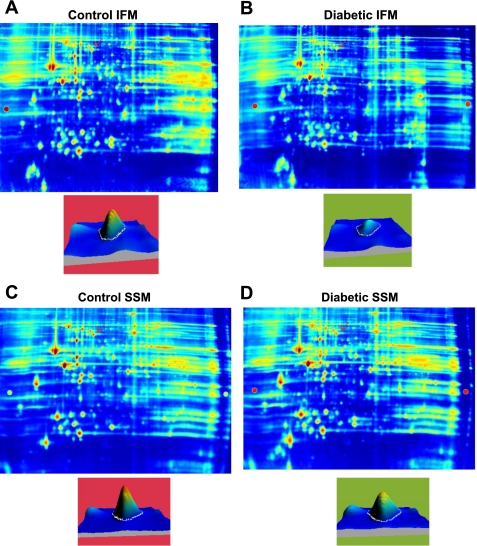
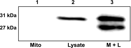
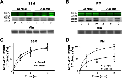
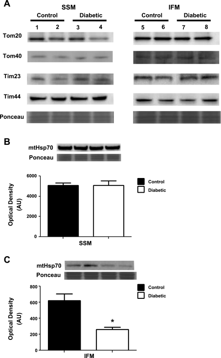
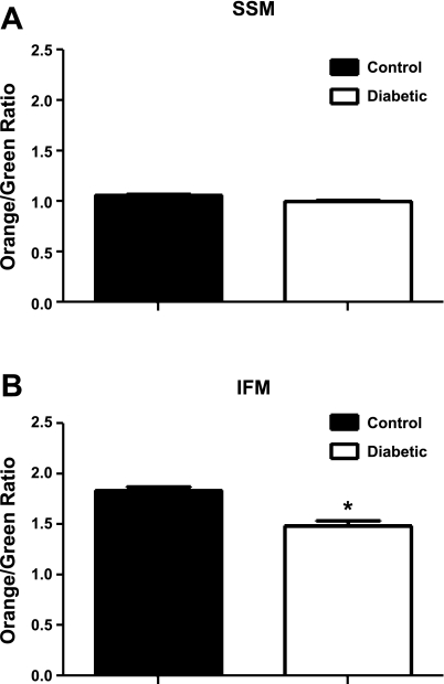
Diabetic cardiomyopathy is associated with increased risk of heart failure in type 1 diabetic patients. Mitochondrial dysfunction is suggested as an underlying contributor to diabetic cardiomyopathy. Cardiac mitochondria are characterized by subcellular spatial locale, including mitochondria located beneath the sarcolemma, subsarcolemmal mitochondria (SSM), and mitochondria situated between the myofibrils, interfibrillar mitochondria (IFM). The goal of this study was to determine whether type 1 diabetic insult in the heart influences proteomic make-up of spatially distinct mitochondrial subpopulations and to evaluate the role of nuclear encoded mitochondrial protein import. Utilizing multiple proteomic approaches (iTRAQ and two-dimensional-differential in-gel electrophoresis), IFM proteomic make-up was impacted by type 1 diabetes mellitus to a greater extent than SSM, as evidenced by decreased abundance of fatty acid oxidation and electron transport chain proteins. Mitochondrial phosphate carrier and adenine nucleotide translocator, as well as inner membrane translocases, were decreased in the diabetic IFM (P < 0.05 for both). Mitofilin, a protein involved in cristae morphology, was diminished in the diabetic IFM (P < 0.05). Posttranslational modifications, including oxidations and deamidations, were most prevalent in the diabetic IFM. Mitochondrial heat shock protein 70 (mtHsp70) was significantly decreased in diabetic IFM (P < 0.05). Mitochondrial protein import was decreased in the diabetic IFM with no change in the diabetic SSM (P < 0.05). Taken together, these results indicate that mitochondrial proteomic alterations in the type 1 diabetic heart are more pronounced in the IFM. Further, proteomic alterations are associated with nuclear encoded mitochondrial protein import dysfunction and loss of an essential mitochondrial protein import constituent, mtHsp70, implicating this process in the pathogenesis of the diabetic heart.


Figures

Fig. 1.
Fig. 1.
Representative iTRAQ spectra. Isolated mitochondrial subpopulations from control and diabetic hearts were labeled with iTRAQ reagents 114 [diabetic subsarcolemmal mitochondria (SSM)], 115 [diabetic interfibrillar mitochondria (IFM)], 116 (control SSM), and 117 (control IFM); then, they were combined for analysis with mass spectrometry. A: representative spectra of simultaneous quantitation of a mtHsp70 peptide in control and diabetic mitochondrial subpopulations. B: MS/MS spectra for the reporter groups of the iTRAQ reagents (114, 115, 116, and 117) from a mtHsp70 peptide. These spectra were used along with other peptides to simultaneously quantify mtHsp70 control and diabetic mitochondrial subpopulations.
Fig. 2.
Fig. 2.
Representative two-dimensional-differential in-gel electrophoresis (2D-DIGE) gels. Samples were labeled with either Cy3 or Cy5, alternating between control and diabetic SSM and IFM. A control and treated mitochondrial sample was run on the same gel, and an internal standard was labeled with Cy2 and run on every gel for gel-to-gel comparisons. A: representative gel showing Cy3-labeled control IFM. B: same gel as A showing Cy5-labeled diabetic IFM. C: representative gel showing Cy3-labeled control SSM. D: same gel as C showing Cy5-labeled control SSM. Below the gel images are individual spots that were identified as mtHsp70 (indicated by the red arrow), and quantitative differences were assessed by examination of peak density.
Fig. 3.
Fig. 3.
MITOGFP1 protein import. Western blot analysis of MitoGFP1 protein import into isolated cardiac mitochondria probed for AcGFP in samples containing 40 μg of mitochondria only (lane 1), 0.5 μl of MitoGFP1 protein lysate (lane 2), or combined mitochondria and lysate (3 μl) (lane 3). The upper 31-kDa band represents the full-length precursor MitoGFP1 in transit and yet to be processed, while the lower 27-kDa band represents a mature cleaved MitoGFP1 protein residing within the mitochondrial matrix.
Fig. 4.
Fig. 4.
Mitochondrial protein import. Effect of type 1 diabetes mellitus on MitoGFP1 import in SSM and IFM subpopulations. Cardiac mitochondrial subpopulations from control and diabetic mice were isolated and incubated with 3 μl of MitoGFP1 protein lysate at 2-, 5-, and 10-min time points. Representative Western blots and fluorescent images from SSM (A) and IFM (B) protein import assay. Graphical representation of mitochondrial protein import performed in SSM (C) and IFM (D) control and diabetic subpopulations. The relative amount of imported MitoGFP1 was determined by densitometry. Values were calculated based upon a 100% scale, and presented as mean ± SE; *P < 0.05 for control vs. diabetic subpopulations. Control for protein loading was confirmed by Ponceau staining; n = 8 per group.
Fig. 5.
Fig. 5.
Western blot analysis of protein import constituents. Key proteins involved in mitochondrial protein import were assessed using Western blot analysis. A: control SSM (lanes 1 and 2), diabetic SSM (lanes 3 and 4), control IFM (lanes 5 and 6), and diabetic IFM (lanes 7 and 8) were analyzed for protein expression in translocases of the outer mitochondrial membrane (Tom20 and Tom40) and inner mitochondrial membrane (Tim23 and Tim44). Representative Western blots and densitometry analysis for mtHsp70 protein expression in SSM (B), and IFM (C) from control and diabetic hearts. Values are presented as means ± SE; *P < 0.05 for control vs. diabetic. Control for protein loading was confirmed with Ponceau staining; n = 5 for each group.
Fig. 6.
Fig. 6.
Mitochondrial subpopulation membrane potential. Isolated mitochondrial subpopulations were incubated with JC-1, and 100,000 gated events were analyzed per sample. Control and diabetic SSM (A) and IFM (B) samples were analyzed. Values are represented as means ± SE. *P < 0.05 for control vs. diabetic; n = 4 for each group.

Comment in

References

    1. Artal-Sanz M, Tavernarakis N. Prohibitin and mitochondrial biology. Trends Endocrinol Metab 20: 394–401, 2009 - PubMed
    1. Baynes JW. Role of oxidative stress in development of complications in diabetes. Diabetes 40: 405–412, 1991 - PubMed
    1. Bradford MM. A rapid and sensitive method for the quantitation of microgram quantities of protein utilizing the principle of protein-dye binding. Anal Biochem 72: 248–254, 1976 - PubMed
    1. Bugger H, Boudina S, Hu XX, Tuinei J, Zaha VG, Theobald HA, Yun UJ, McQueen AP, Wayment B, Litwin SE, Abel ED. Type 1 diabetic akita mouse hearts are insulin sensitive but manifest structurally abnormal mitochondria that remain coupled despite increased uncoupling protein 3. Diabetes 57: 2924–2932, 2008 - PMC - PubMed
    1. Bugger H, Chen D, Riehle C, Soto J, Theobald HA, Hu XX, Ganesan B, Weimer BC, Abel ED. Tissue-specific remodeling of the mitochondrial proteome in type 1 diabetic akita mice. Diabetes 58: 1986–1997, 2009 - PMC - PubMed

Publication types

MeSH terms