. 2010 Nov 23;107(47):20441-6.
doi: 10.1073/pnas.1013827107. Epub 2010 Nov 3.

Antibodies protect against intracellular bacteria by Fc receptor-mediated lysosomal targeting

Affiliations

Antibodies protect against intracellular bacteria by Fc receptor-mediated lysosomal targeting

Nicole Joller et al. Proc Natl Acad Sci U S A. .

Abstract

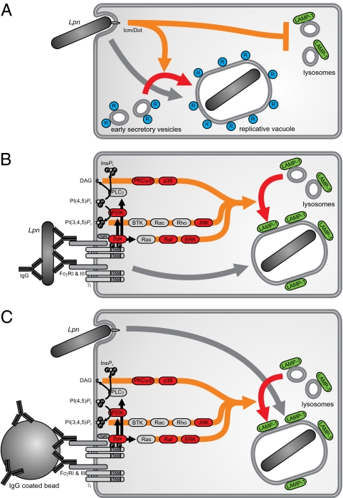
The protective effect of antibodies (Abs) is generally attributed to neutralization or complement activation. Using Legionella pneumophila and Mycobacterium bovis bacillus Calmette-Guérin as a model, we discovered an additional mechanism of Ab-mediated protection effective against intracellular pathogens that normally evade lysosomal fusion. We show that Fc receptor (FcR) engagement by Abs, which can be temporally and spatially separated from bacterial infection, renders the host cell nonpermissive for bacterial replication and targets the pathogens to lysosomes. This process is strictly dependent on kinases involved in FcR signaling but not on host cell protein synthesis or protease activation. Based on these findings, we propose a mechanism whereby Abs and FcR engagement subverts the strategies by which intracellular bacterial pathogens evade lysosomal degradation.


Conflict of interest statement

The authors declare no conflict of interest.

Figures

Fig. 1.
Fig. 1.
Antibodies protect from Lpn infection and inhibit Lpn growth by redirection into lysosomes in vitro. (A) B6 mice were vaccinated with Lpn (memory) or left naive. Colony-forming units (CFU) were determined in the lung and BAL 2 d after i.n. challenge with IgG- or IgA-opsonized Lpn (IgG and IgA) or untreated Lpn (naive; n = 3). (B–D) RAW MΦ were infected with Lpn pretreated with medium, naive serum, or Lpn-specific immune serum, and (B) intracellular growth of Lpn was measured. (C and D) LCVs were isolated from infected MΦ and stained for the T4SS substrate SidC (blue) and the lysosomal associated membrane protein 1 (LAMP-1) (red), and the percentage of Lpn (green) localizing in LAMP-1+ vacuoles was quantified. T4SS-deficient Lpn (ΔT), which are unable to evade lysosomal fusion, were used as positive controls for the localization in LAMP-1+ vacuoles. Dotted lines visualize LCVs. (Scale bar: 2 μm.) At least 40 LCVs were analyzed in each experimental condition. SDs from three independent experiments are shown. (E and F) Bone marrow-derived MΦ from B6 or FcRγ−/− mice were infected with Lpn pretreated with medium, naive serum, Lpn-specific immune serum, or Lpn-specific F(ab′)2. LCVs were isolated from infected MΦ and stained for the lysosomal marker LAMP-1 (red), and the percentage of Lpn (green) localized in LAMP-1+ vacuoles was quantified. Dotted lines visualize LCVs. (Scale bar: 2 μm.) At least 40 LCVs were analyzed in each experimental condition, and SDs from three independent experiments are shown. Means ± SD are indicated. P values were calculated by Student t test (two-tailed, unpaired). ns, not significant.
Fig. 2.
Fig. 2.
Host FcR and the Fc portion of Abs are essential for protection from Lpn in vivo. B6 mice were vaccinated with Lpn (memory) or were left naive. (A) B6 or FcRγ−/− mice were challenged i.n. with IgG- or (B) F(ab′)2-opsonized Lpn (IgG, F(ab′)2) or untreated Lpn (naive, memory), and CFUs were determined in the lung and BAL 2 d later. Means ± SD are indicated (n = 3). P values were calculated by Student t test (two-tailed, unpaired). ns, not significant.
Fig. 3.
Fig. 3.
FcR activation and downstream signaling are essential for directing Lpn into lysosomes in vitro. (A) RAW MΦ were incubated with IgG- (red ●) or MSA-beads (green ○) or were left untreated (squares). Cells were then infected with wt (■, red ●, and green ○) or ΔT Lpn (□), and intracellular growth was measured. (B and C) RAW MΦ expressing LAMP-1–GFP were pretreated with FcR signaling inhibitors or left untreated, and they were subsequently incubated with IgG- or MSA-beads or media. The cells were then infected with DsRed-expressing wt or ΔT Lpn, and the percentage of bacteria localizing in LAMP-1+ vacuoles was quantified. (Scale bar: B, 5 μm.) (C) One hundred LCVs were analyzed in each experimental condition, and SDs from two to four independent experiments are shown. (D) RAW MΦ expressing LAMP-1–GFP were pretreated with PKC inhibitors or left untreated and then were infected with DsRed-expressing wt or ΔT Lpn pretreated with medium, naive serum, or Lpn-specific immune serum; the percentage of bacteria localizing in LAMP-1+ vacuoles was quantified. One hundred LCVs were analyzed in each experimental condition, and SDs from three to five independent experiments are shown. P values were calculated by Student t test (two-tailed, unpaired). ns, not significant.
Fig. 4.
Fig. 4.
Abs redirect Lpn to lysosomes in vivo. B6 mice were vaccinated with Lpn (memory) or were left naive. The mice were challenged i.n. with IgG- or F(ab′)2-opsonized (IgG, F(ab′)2) or untreated Lpn GFP (naive, memory), and the localization of the bacteria was analyzed 16–20 h after infection. Lungs were stained for the lysosomal marker LAMP-1 (red), actin (blue), and nuclei (white), and the percentage of Lpn (green) localizing in LAMP-1+ compartments was quantified. (Scale bar: A, 20 μm.) (B) n = 20 each; means ± SD are depicted. P values were calculated by Student t test (two-tailed, unpaired). ns, not significant.
Fig. 5.
Fig. 5.
The relevance of the FcR trigger and its downstream signaling for the intracellular fate of Lpn in host cells. Schematic view of Lpn taken up by a MΦ. (A) Using their T4SS, the bacteria actively avoid fusion with lysosomes but acquire vesicles trafficking between the endoplasmic reticulum (ER) and Golgi, and finally, they replicate in a cellular compartment studded with ribosomes. (B) Opsonized bacteria activate FcRs and are subsequently targeted to lysosomal compartments. (C) Temporal and spatial separation of the FcR activation and FcR-independent uptake of Lpn into the MΦ also result in lysosomal targeting of the bacteria.

Similar articles

Cited by

References

    1. Flannagan RS, Cosío G, Grinstein S. Antimicrobial mechanisms of phagocytes and bacterial evasion strategies. Nat Rev Microbiol. 2009;7:355–366. - PubMed
    1. Vergne I, et al. Mechanism of phagolysosome biogenesis block by viable Mycobacterium tuberculosis. Proc Natl Acad Sci USA. 2005;102:4033–4038. - PMC - PubMed
    1. Clemens DL, Lee BY, Horwitz MA. Deviant expression of Rab5 on phagosomes containing the intracellular pathogens Mycobacterium tuberculosis and Legionella pneumophila is associated with altered phagosomal fate. Infect Immun. 2000;68:2671–2684. - PMC - PubMed
    1. McDade JE, et al. Legionnaires’ disease: Isolation of a bacterium and demonstration of its role in other respiratory disease. N Engl J Med. 1977;297:1197–1203. - PubMed
    1. Vogel JP, Andrews HL, Wong SK, Isberg RR. Conjugative transfer by the virulence system of Legionella pneumophila. Science. 1998;279:873–876. - PubMed

Publication types