. 2010 Dec 1;185(11):6819-30.
doi: 10.4049/jimmunol.1000448. Epub 2010 Nov 3.

Re-examination of CD91 function in GRP94 (glycoprotein 96) surface binding, uptake, and peptide cross-presentation

Affiliations

Re-examination of CD91 function in GRP94 (glycoprotein 96) surface binding, uptake, and peptide cross-presentation

Angela R Jockheck-Clark et al. J Immunol. .

Abstract

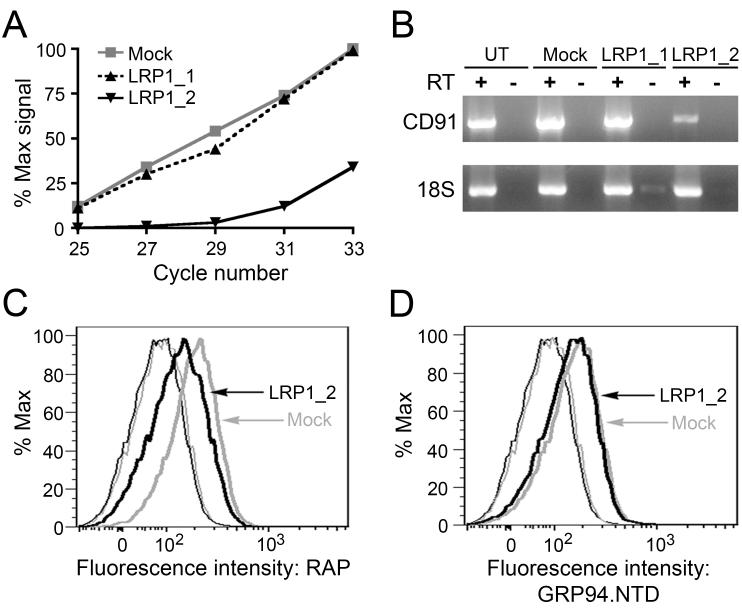
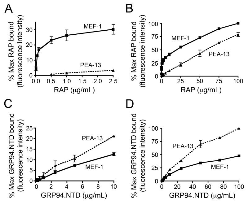
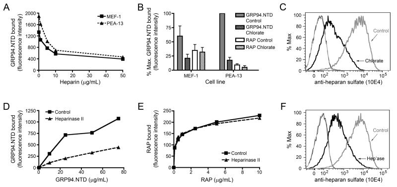
GRP94 (gp96)-peptide complexes can be internalized by APCs and their associated peptides cross-presented to yield activation of CD8(+) T cells. Investigations into the identity (or identities) of GRP94 surface receptors have yielded conflicting results, particularly with respect to CD91 (LRP1), which has been proposed to be essential for GRP94 recognition and uptake. To assess CD91 function in GRP94 surface binding and endocytosis, these parameters were examined in mouse embryonic fibroblast (MEF) cell lines whose expression of CD91 was either reduced via RNA interference or eliminated by genetic disruption of the CD91 locus. Reduction or loss of CD91 expression abrogated the binding and uptake of receptor-associated protein, an established CD91 ligand. Surface binding and uptake of an N-terminal domain of GRP94 (GRP94.NTD) was unaffected. GRP94.NTD surface binding was markedly suppressed after treatment of MEF cell lines with heparin, sodium chlorate, or heparinase II, demonstrating that heparin sulfate proteoglycans can function in GRP94.NTD surface binding. The role of CD91 in the cross-presentation of GRP94-associated peptides was examined in the DC2.4 dendritic cell line. In DC2.4 cells, which express CD91, GRP94.NTD-peptide cross-presentation was insensitive to the CD91 ligands receptor-associated protein or activated α(2)-macroglobulin and occurred primarily via a fluid-phase, rather than receptor-mediated, uptake pathway. These data clarify conflicting data on CD91 function in GRP94 surface binding, endocytosis, and peptide cross-presentation and identify a role for heparin sulfate proteoglycans in GRP94 surface binding.


Figures

Figure 1
Figure 1. Expression and biophysical characterization of GRP94.NTD
Recombinant GRP94.LREK was expressed, purified, and depyrogenated. A. 5 μg GRP94.LREK (69-337) (lane 2) was resolved by 12.5% SDS-PAGE gel and detected by Coomassie Blue staining. B. To examine the oligomeric state of purified GRP94.LREK, 7.5 μg GRP94.LREK was resolved by 6% native PAGE and detected by Coomassie Blue staining (lane 1). Incubation at 42°C for 30 min promotes the formation of higher-order GRP94.LREK oligomers (lane 2). C. Analytical ultracentrifugation was performed on 2.3 mg/mL GRP94.LREK at 4°C. The protein displayed a high degree of homogeneity, and mass average calculations yielded a molecular mass estimation of 41 kDa.
Figure 2
Figure 2. Suppression of CD91 expression does not reduce GRP94.NTD surface binding to MEF-1 cells
MEF-1 cells were transiently transfected with either of two CD91-targeting siRNAs (LRP1_1 and LRP1_2) or a vector only (mock) control, and examined for changes in CD91 expression 24 h post-transfection. A. Total RNA samples were isolated and CD91 mRNA knockdown efficiency was determined by RT-PCR. CD91 cDNA from mock-transfected cells displayed linear amplification between PCR cycles 25 and 33 (R2 = 0.9974). B. Untransfected (UT), mock-transfected, and siRNA-transfected cells were examined for changes in CD91 mRNA levels using RT-PCR. All samples were tested for DNA contamination using a paired transcriptase-deficient (RT-) reaction, and were examined for off-target effects to 18S rRNA. C & D. MEF-1 cells were transfected with LRP1_2 siRNA or a mock control and analyzed by flow cytometry. CD91 surface expression was determined using 10 μg/mL RAP (C), and GRP94.NTD surface binding was conducted with 25 μg/mL GRP94.NTD (D): unstained cells (thin lines), stained cells (bold lines), mock-transfected cells (grey lines), and LRP1_2 siRNA transfected cells (black lines). Mock-transfected and untransfected cells displayed identical RAP and GRP94.NTD binding (data not shown). The data presented are representative of three independent replicates.
Figure 3
Figure 3. GRP94.NTD binds MEF-1 and PEA-13 cells independently of CD91 expression
A. MEF-1 and PEA-13 cells were analyzed for CD91 expression using RT-PCR. All samples were tested for DNA contamination using a paired transcriptase-deficient (RT-) reaction, and were examined for off-target effects to 18S rRNA. B & C. MEF-1 and PEA-13 cells were incubated with 10 μg/mL RAP (B) or 25 μg/mL GRP94.NTD (C), washed, and analyzed by flow cytometry: unstained cells (thin lines), stained cells (bold lines), MEF-1 cells (black lines), and PEA-13 cells (grey lines). D. MEF-1 cells were incubated with 10μg/mL RAP in the absence or presence of 100-fold molar excess unlabeled RAP, GRP94.NTD, or BSA. Cells were then washed and analyzed by flow cytometry: unstained cells (thin grey line), RAP (bold black line), unlabeled RAP (thin black line), unlabeled GRP94.NTD (bold grey line), and unlabeled BSA (shaded grey line). Data presented are representative of three independent replicates.
Figure 4
Figure 4. Binding characteristics of RAP and GRP94.NTD to MEF-1 and PEA-13 cells
MEF-1 and PEA-13 cells were incubated with increasing concentrations of RAP (A and B) or GRP94.NTD (C and D). Cells were subsequently washed and analyzed by flow cytometry. Results are expressed as the mean of three independent experiments ± SD.
Figure 5
Figure 5. Modulation of cell surface HSPG structures decreases GRP94.NTD surface binding
A. MEF-1 and PEA-13 cells were incubated with increasing concentrations of heparin, washed, incubated with 25 μg/mL GRP94.NTD, and analyzed by flow cytometry. B. MEF-1 and PEA-13 cells were grown in the absence (solid) or presence (striped) of 20 mM sodium chlorate. Cells were incubated with 25 μg/mL GRP94.NTD (grey) or 10 μg/mL RAP (white), washed, and analyzed by flow cytometry. Results are expressed as the mean of three independent experiments ± SD. C. To confirm that sodium chlorate treatment reduced HSPG sulfation, MEF-1 cells were stained with the anti-heparan sulfate antibody 10E4, washed, and analyzed by flow cytometry: unstained cells (thin lines), stained cells (bold lines), control cells (grey lines), and chlorate-treated cells (black lines). D & E. MEF-1 cells were incubated with or without 0.01 IU/mL heparinase II (Hep’ase) for 90 min at 37°C. Cells were then incubated with increasing concentrations of GRP94.NTD (D) or RAP (E), and analyzed by flow cytometry. F. To confirm efficient de-sulfation by heparinase II, MEF-1 cells were stained with the anti-heparan sulfate antibody 10E4, washed, and analyzed by flow cytometry: unstained cells (thin lines), stained cells (bold lines), control cells (grey lines), and chlorate-treated cells (black lines). All data presented are representative of at least three independent replicates.
Figure 6
Figure 6. GRP94.NTD and RAP are internalized via spatially and kinetically distinct pathways
A & B. MEF-1 cells were incubated with either 10 μg/mL RAP or 25 μg/mL GRP94.NTD, washed, warmed to 37°C for 0, 5, 20, or 60 min, and placed on ice to arrest endocytosis. Residual surface-bound ligands were removed by proteolysis on ice. The percent protease-resistant signal was calculated by normalizing the protease resistant signal to the total signal at each time point. Results are expressed as the mean of three independent experiments ± SD B. Comparison of MEF-1 cells treated either as in A (control) or treated with 10 μg/mL RAP prior to GRP94.NTD incubation and excess RAP during internalization (RAP Competition). Residual surface-bound ligands were removed by proteolysis on ice. The dashed gray bar indicates total GRP94.NTD surface binding prior to treatment with extracellular protease. C-F. MEF-1 cells were incubated with 5 μg/mL RAP (red) and 40 μg/mL GRP94.NTD (blue), washed, and warmed to 37°C for 0 (C), 5 (D), 15 (E), or 60 (F) min to allow internalization. Cells were then fixed and processed for confocal microscopy. Scale bars: 20 μm. All data presented are representative of three independent replicates.
Figure 7
Figure 7. Proteolysis enhances the fluorescence yield of fluor-conjugated GRP94.NTD
A & B. Fluor-conjugated GRP94.NTD was incubated with trypsin at room temperature. At various time points, the reaction was quenched by addition of TCA and precipitated protein was analyzed by SDS-PAGE and Coomassie Blue staining (A). Values were normalized to the untreated control (lane 1). GRP94.NTD fluorescence was determined at thirty second intervals (B). Relative GRP94 fluorescence was normalized to the point of highest fluorescence intensity. C & D. MEF-1 cells were pre-incubated in the absence or presence of 100 μg/mL leupeptin. Cells were then incubated with 25 μg/mL GRP94.NTD on ice, washed, and warmed to 37°C for 0 (bold black line) or 60 min in the absence (dashed line) or presence (bold grey line) of 100 μg/mL leupeptin. D. Data from C represented as a bar graph.
Figure 8
Figure 8. GRP94.NTD internalization and processing efficiencies are independent of exogenous CD91 ligands
A & B. DC2.4 cells were incubated with increasing concentrations of RAP (A) or GRP94.NTD (B). Cells were subsequently washed and analyzed by flow cytometry. Results are expressed as the mean of two independent experiments ± SD. C. DC2.4 cells were incubated with 10 μg/mL GRP94.NTD in the absence or presence of 500 μg/mL RAP, 300 μg/mL α2M*, or 300 μg/mL native α2M for 30 min on ice. Cells were then washed, and analyzed by flow cytometry. D. DC2.4 cells were prepared as in C, and then incubated in the absence or presence of 500 μg/mL RAP, 300 μg/mL α2M*, or 300 μg/mL native α2M for 60 min at 37°C. E. DC2.4 cells were incubated with media only, 1 μM free ova20 peptide, uncomplexed GRP94.NTD, or increasing concentration of GRP94.NTD/ova20 complex. Cells were then incubated for 4 h at 37°C, washed, and incubated with OT-1 splenocytes for 24 h. IFN-γ secretion was quantified by ELISA. F. DC2.4 cells were incubated with 10 μg/mL GRP94.NTD/ova20 complex in the absence or presence of 500 μg/mL RAP, 300 μg/mL α2M*, 300 μg/mL native α2M, or 500 μg/mL uncompled GRP94.NTD, and treated as in E. G. DC2.4 cells were incubated with 10 μg/mL RAP in the absence or presence of 100 μg/mL unlabeled RAP for 5 min. H. DC2.4 cells were incubated with 10 μg/mL GRP94.NTD in the absence or presence of 250 μg/mL GRP94.NTD or 100 μg/mL RAP for 20 min. Cells were then placed on ice to arrest endocytosis, rinsed with cold buffer, and analyzed by flow cytometry. Samples were normalized to their respective no competitor controls. Results are expressed as the mean of two independent experiments ± SD.

Similar articles

Cited by

References

    1. Srivastava P. Roles of heat-shock proteins in innate and adaptive immunity. Nat Rev Immunol. 2002;2:185–194. - PubMed
    1. Nicchitta CV. Re-evaluating the role of heat-shock protein-peptide interactions in tumour immunity. Nat Rev Immunol. 2003;3:427–432. - PubMed
    1. Binder RJ, Harris ML, Menoret A, Srivastava PK. Saturation, competition, and specificity in interaction of heat shock proteins (hsp) gp96, hsp90, and hsp70 with CD11b+ cells. J Immunol. 2000;165:2582–2587. - PubMed
    1. Wassenberg JJ, Dezfulian C, Nicchitta CV. Receptor mediated and fluid phase pathways for internalization of the ER Hsp90 chaperone GRP94 in murine macrophages. J Cell Sci. 1999;112(Pt 13):2167–2175. - PubMed
    1. Arnold-Schild D, Hanau D, Spehner D, Schmid C, Rammensee HG, de la Salle H, Schild H. Cutting edge: receptor-mediated endocytosis of heat shock proteins by professional antigen-presenting cells. J Immunol. 1999;162:3757–3760. - PubMed

Publication types

MeSH terms