

Cortical and thalamic innervation of direct and indirect pathway medium-sized spiny neurons in mouse striatum
- PMID: 21048118
- PMCID: PMC6633626
- DOI: 10.1523/JNEUROSCI.1623-10.2010
Cortical and thalamic innervation of direct and indirect pathway medium-sized spiny neurons in mouse striatum
Abstract
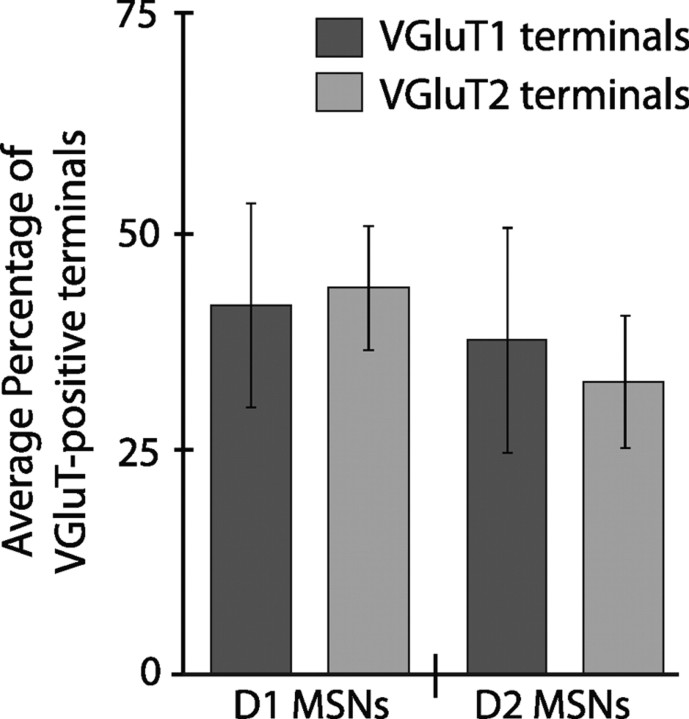
The striatum receives major excitatory inputs from the cortex and thalamus that predominantly target the spines of medium-sized spiny neurons (MSNs). We aimed to determine whether there is any selectivity of these two excitatory afferents in their innervation of direct and indirect pathway MSNs. To address this, we used bacterial artificial chromosome transgenic mice, in which enhanced green fluorescent protein (EGFP) reports the presence of D(1) or D(2) dopamine receptor subtypes, markers of direct and indirect pathway MSNs, respectively. Excitatory afferents were identified by the selective expression of vesicular glutamate transporter type 1 (VGluT1) by corticostriatal afferents and vesicular glutamate transporter type 2 (VGluT2) by thalamostriatal afferents. A quantitative electron microscopic analysis was performed on striatal tissue from D(1) and D(2) mice that was double immunolabeled to reveal the EGFP and VGluT1 or VGluT2. We found that the proportion of synapses formed by terminals derived from the cortex and thalamus was similar for both direct and indirect pathway MSNs. Furthermore, qualitative analysis revealed that individual cortical or thalamic terminals form synapses with both direct and indirect pathway MSNs. Similarly, we observed a convergence of cortical and thalamic inputs onto individual MSNs of both direct and indirect pathway: individual EGFP-positive structures received input from both VGluT2-positive and VGluT2-negative terminals. These findings demonstrate that direct and indirect pathway MSNs are similarly innervated by cortical and thalamic afferents; both projections are thus likely to be critical in the control of MSNs and hence play fundamental roles in the expression of basal ganglia function.
Figures


Similar articles
-
Convergence of cortical and thalamic input to direct and indirect pathway medium spiny neurons in the striatum.Brain Struct Funct. 2014 Sep;219(5):1787-800. doi: 10.1007/s00429-013-0601-z. Epub 2013 Jul 6. Brain Struct Funct. 2014. PMID: 23832596 Free PMC article.
-
Homeostatic regulation of excitatory synapses on striatal medium spiny neurons expressing the D2 dopamine receptor.Brain Struct Funct. 2016 May;221(4):2093-107. doi: 10.1007/s00429-015-1029-4. Epub 2015 Mar 18. Brain Struct Funct. 2016. PMID: 25782435
-
Differential synaptology of vGluT2-containing thalamostriatal afferents between the patch and matrix compartments in rats.J Comp Neurol. 2006 Nov 10;499(2):231-43. doi: 10.1002/cne.21099. J Comp Neurol. 2006. PMID: 16977615 Free PMC article.
-
Postsynaptic integration of glutamatergic and dopaminergic signals in the striatum.Prog Neurobiol. 1994 Oct;44(2):163-96. doi: 10.1016/0301-0082(94)90037-x. Prog Neurobiol. 1994. PMID: 7831476 Review.
-
Excitatory extrinsic afferents to striatal interneurons and interactions with striatal microcircuitry.Eur J Neurosci. 2019 Mar;49(5):593-603. doi: 10.1111/ejn.13881. Epub 2018 Mar 25. Eur J Neurosci. 2019. PMID: 29480942 Free PMC article. Review.
Cited by
-
Altered mGluR5-Homer scaffolds and corticostriatal connectivity in a Shank3 complete knockout model of autism.Nat Commun. 2016 May 10;7:11459. doi: 10.1038/ncomms11459. Nat Commun. 2016. PMID: 27161151 Free PMC article.
-
Comparative Ultrastructural Analysis of Thalamocortical Innervation of the Primary Motor Cortex and Supplementary Motor Area in Control and MPTP-Treated Parkinsonian Monkeys.Cereb Cortex. 2021 Jun 10;31(7):3408-3425. doi: 10.1093/cercor/bhab020. Cereb Cortex. 2021. PMID: 33676368 Free PMC article.
-
Action selection performance of a reconfigurable basal ganglia inspired model with Hebbian-Bayesian Go-NoGo connectivity.Front Behav Neurosci. 2012 Oct 2;6:65. doi: 10.3389/fnbeh.2012.00065. eCollection 2012. Front Behav Neurosci. 2012. PMID: 23060764 Free PMC article.
-
Thalamo-Nucleus Accumbens Projections in Motivated Behaviors and Addiction.Front Syst Neurosci. 2021 Jul 15;15:711350. doi: 10.3389/fnsys.2021.711350. eCollection 2021. Front Syst Neurosci. 2021. PMID: 34335197 Free PMC article. Review.
-
Chronic and Acute Manipulation of Cortical Glutamate Transmission Induces Structural and Synaptic Changes in Co-cultured Striatal Neurons.Front Cell Neurosci. 2021 Feb 18;15:569031. doi: 10.3389/fncel.2021.569031. eCollection 2021. Front Cell Neurosci. 2021. PMID: 33679324 Free PMC article.
References
-
- Barroso-Chinea P, Castle M, Aymerich MS, Lanciego JL. Expression of vesicular glutamate transporters 1 and 2 in the cells of origin of the rat thalamostriatal pathway. J Chem Neuroanat. 2008;35:101–107. - PubMed
-
- Bennett BD, Bolam JP. Synaptic input and output of parvalbumin-immunoreactive neurons in the neostriatum of the rat. Neuroscience. 1994;62:707–719. - PubMed
-
- Bolam JP, editor. Experimental neuroanatomy. Oxford: Oxford UP; 1992.
Publication types
MeSH terms
Substances
Grants and funding
LinkOut - more resources
Full Text Sources
Molecular Biology Databases