. 2010 Nov 3;30(44):14735-44.
doi: 10.1523/JNEUROSCI.2919-10.2010.

Dynamic plasticity of axons within a cutaneous milieu

Affiliations

Dynamic plasticity of axons within a cutaneous milieu

Chu Cheng et al. J Neurosci. .

Abstract

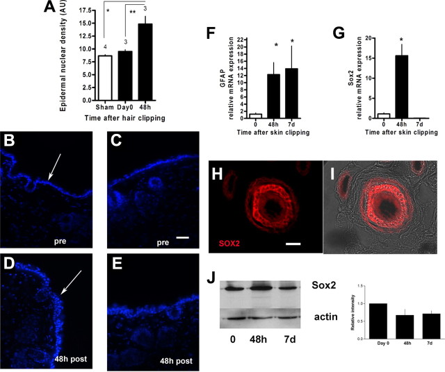
The skin is a repository of sensory axons immersed within the turnover of epidermal, follicular, and dermal cellular constituents. We show that epidermal and perifollicular axons within intact hairy skin of mice possess a remarkable dynamic plasticity linked to their microenvironment. For example, the majority of epidermal axons express the growth protein GAP43. Unexpectedly, we induced new cutaneous axogenesis by simple and noninvasive hair clipping, a response linked to a series of changes in their cutaneous neighbors. In thy-1 YFP transgenic mice with fluorescent axons, superficial epidermal and perifollicular cells newly acquired YFP, indicating diffuse activation by clipping despite the absence of skin injury. At 48 h after clipping, this activation was accompanied by a rise in the number of epidermal cells, transient rises in mRNA of Sox2, a marker of follicular stem cells, and a rise in mRNA of glial fibrillary acidic protein, a marker of glial cells. Axons responded with rises in their numbers in the epidermis and around dermal hair follicles. Linking these responses were early, large, and selective rises in hepatic growth factor (HGF) mRNA, with its protein identified in epidermal cells, perifollicular cells, and sensory axons. Moreover, these elements also expressed the HGF receptor c-Met, especially in small caliber sensory neurons. Finally, we identified concurrent rises in Rac1 activation, a downstream target of ligated c-Met. Together, these results confirm critical linkages between sensory axons and their cutaneous milieu. We believe that the plasticity is provoked by follicular-originating cutaneous activation with HGF and Rac1 signaling, allowing cross talk and axonal remodeling.


Figures

Figure 1.
Figure 1.
Intact skin has evidence of inherent plasticity. AC, Images of dorsal trunk skin of the mouse labeled with antibodies directed against PGP9.5, which is expressed in all dermal and epidermal fibers. Note the large caliber nerve bundles within the dermis that ramify outward into the epidermis. The epidermal profiles are highly tortuous. The arrow points to a dermal hair follicle, just below the epidermis (central shaft area is dark), that is associated with axons. DF, Axons labeled for GAP43/B50, a regeneration-related growth molecule, are observed in large numbers of dermal (including perifollicular axons; D, arrow) and epidermal axons indicating an ongoing plastic, regenerative phenotype. Note the abrupt change in trajectory in a GAP43/B50-labeled epidermal axon (E′, arrow). GI, Dermal axons (PGP9.5) are closely accompanied (arrows) by fine activated glial cell processes (GFAP), indicating a close and potentially supportive role for axon regeneration. Asterisks indicates outer surface of the skin and arrowheads indicate the dermal–epidermal junction. Scale bar, 20 μm.
Figure 2.
Figure 2.
Hair clipping alters thy-1 YFP expression in perifollicular cells without damaging the skin. A, Keratin-15 mRNA expression is unchanged or reduced, rather than elevated, following noninvasive skin clipping (48 h, 7 d clp), indicating that deeper epidermal layers were not injured. In contrast, skin having undergone an invasive biopsy wound (7 d bx) had a rise in keratin-15 expression (*p = 0.05, ANOVA). BE, In percutaneous images from live mice, the same area of skin in the same mouse is seen by epifluorescence before (B, D) and after (C, E) clipping. Note the donut shaped (E, arrow; hair shaft is in the center) intense areas of YFP expression 7 d following a single episode of hair clipping at low (C) and high (E) magnification. The expression is often diffuse and not confined to discrete profiles (arrowhead). Scale bar: D, E, 200 μm; B, C, 500 μm. F, G, Identification of YFP-expressing axons in the skin is shown using conventional epifluorescence (F) and confocal serial imaging (G) of the skin in live mice 7 d following shaving. Scale bar,100 μm.
Figure 3.
Figure 3.
Cutaneous plasticity develops rapidly following noninvasive hair clipping. A, Density of epidermal nuclei per unit area in the epidermis. There is a significant rise in epidermal nuclear density 48 h following hair clipping compared with a sham intervention (*p < 0.01) or with d 0 (**p < 0.01). Sham clipping involved applying the clipper to the skin without powering the blades (one-way ANOVA, p = 0.002; post hoc Tukey test, n = 3 on d 0, n = 3 at 48 h, n = 4 for sham). BE, Examples of epidermal nuclear labeling (B and C, without previous clipping; D and E, 48 h following clipping). The arrows indicate epidermal nuclear labeling and the epidermis faces upward. F, G, Clipping was also associated with rises in mRNA levels of GFAP expressed in skin glial cells and in mRNA of Sox2 (G; *one-way ANOVA, p = 0.0002; post hoc Tukey 48 h vs d 0, p < 0.05; n = 5 at d 0 and 7, n = 5 at 48 h), an epidermal stem cell marker (F; *one-way ANOVA, p = 0.036; post hoc Tukey d 7 vs d 0, p < 0.05; n = 10 at d 0, n = 6 at 48 h; n = 7 at d 7). H, I, Sox2 was expressed in perifollicular cells, surrounding hair shafts (I, with transmitted light). J, Sox2 expression by immunoblot as indicated, compared with actin loading controls. There was no significant change in its overall protein expression at 48 h or 7 d after skin hair clipping. Scale bars, 50 μm.
Figure 4.
Figure 4.
Cutaneous plasticity is accompanied by axogenesis. A, There were early (48 h) rises in neurofilament heavy unit (Nf200) mRNA (A; *one-way ANOVA, p < 0.0001; post hoc Tukey's t test 0 vs 48 h; p < 0.001; n = 10 at d 0, n = 6 at 48 h, n = 4 at d 7), not observed at the later time point. BE, Representative sections of dorsal cutaneous skin showing rises in epidermal innervation (B, C) and perifollicular innervation (D, E) in controls (B, D) and at 7 d (C, E) following simple hair clipping (red, axons labeled with antibody directed toward PGP9.5, epidermis is directed upward; *outer surface of skin; arrowhead, dermal–epidermal junction). Scale bar: B, C, 20 μm; D, E, 100 μm. F, G, Data showing rises in the density of epidermal vertically oriented axons (F; *one-way ANOVA, p = 0.0004; post hoc Tukey's test d 0 vs 7; p < 0.01; n = 4 for each group) and the percentage of perifollicular axons (G; *one-way ANOVA, p = 0.0016; post hoc Tukey's test d 0 vs 7; p < 0.01; n = 4 for d 0 and 7; n = 5 for 48 h) after hair clipping. HO, Images of dorsal cutaneous skin in mice with a thy-1YFP transgene. The images are triple labeled with DAPI to highlight follicular cell nuclei (H, I, blue), PGP9.5 to identify axons (J, K, green) and anti-GFP (YFP) to enhance the transgene YFP expression (L, M, red) and merged (N, O). The images are sections through the epidermis and dermis in the plane of a telogen hair follicle either immediately after hair clipping (H, J, L, N, d 0) or 7 d following hair clipping in an early anagen phase (I, K, M, O). Clipping left a short shaft of hair in place and did not damage the skin. Note the enhanced innervation of the perifollicular area (arrow points to a perifollicular axon) confirmed to be axons using anti-GFP (YFP) and PGP9.5. *, Outer surface of skin. Scale bar, 50 μm.
Figure 5.
Figure 5.
HGF ligand and receptor are expressed in sensory neurons, axons, epidermal cells, and follicular cells. AG, Immunohistochemistry illustrating expression of c-Met, the HGF receptor in lumbar dorsal root ganglia sensory neurons (AC, C′). Note the lack of colocalization with the neurofilament marker (Nf200) of large neurons and its expression in smaller caliber neurons (arrow). Clusters of small c-Met axons interspaced among large neurofilament labeled axons in the sciatic nerve (D–F) and in small dermal nerve bundles in the dermis (D′, E′). c-Met was also expressed in epidermal cells (G). HO, Data showing mRNA expression of HGF ligand in skin samples, indicating a large rise in mRNA expression at 48 h following hair clipping (H, ANOVA, p < 0.0001; post hoc Tukey's 0 vs 48 h, p < 0.001). HGF was expressed in epidermal cells (I), perifollicular cells, and axons in the dermis (I, arrow shows a cluster of hair follicles; JL, arrow shows a dermal axon associated with hair follicles), and in most sensory neurons in the lumbar dorsal root ganglia (M–O), frequently double labeling with neurofilament. Scale bar: AC, 50 μm; DF, 33 μm; G, I, 100 μm; JL, 21 μm; MO, 50 μm.
Figure 6.
Figure 6.
Rac1 is activated by noninvasive hair clipping. Pull-down assays showing activated Rac1 and total Rac1. Lane 1 is a molecular marker (20 kDa). Lane 2 is from skin immediately after clipping, lane 3 at 48 h following hair clipping, and lane 4 at 7 d following hair clipping. Lane 5 is a positive control (from the manufacturer) and lane 6 is a negative control. Protein from each band was harvested from four mice (two back skin biopsy samples for each mouse), repeated three times in separate groups of mice. Quantitation identifies a significant rise in the ratio of activated Rac1 to total Rac1 at 48 h following shaving (ANOVA, p = 0.02; post hoc Tukey's 0 vs 48 h, p < 0.05).

Similar articles

Cited by

References

    1. Avilion AA, Nicolis SK, Pevny LH, Perez L, Vivian N, Lovell-Badge R. Multipotent cell lineages in early mouse development depend on SOX2 function. Genes Dev. 2003;17:126–140. - PMC - PubMed
    1. Biernaskie JA, McKenzie IA, Toma JG, Miller FD. Isolation of skin-derived precursors (SKPs) and differentiation and enrichment of their Schwann cell progeny. Nat Protoc. 2006;1:2803–2812. - PubMed
    1. Biernaskie J, Paris M, Morozova O, Fagan BM, Marra M, Pevny L, Miller FD. SKPs Derive from hair follicle precursors and exhibit properties of adult dermal stem cells. Cell Stem Cell. 2009;5:610–623. - PMC - PubMed
    1. Botchkarev VA, Paus R. Molecular biology of hair morphogenesis: development and cycling. J Exp Zool B Mol Dev Evol. 2003;298:164–180. - PubMed
    1. Botchkarev VA, Eichmüller S, Johansson O, Paus R. Hair cycle-dependent plasticity of skin and hair follicle innervation in normal murine skin. J Comp Neurol. 1997;386:379–395. - PubMed

Publication types

MeSH terms