Comparative Study
. 2010 Nov 3;30(44):14854-61.
doi: 10.1523/JNEUROSCI.3895-10.2010.

Differential short-term plasticity at convergent inhibitory synapses to the substantia nigra pars reticulata

Affiliations
Comparative Study

Differential short-term plasticity at convergent inhibitory synapses to the substantia nigra pars reticulata

William M Connelly et al. J Neurosci. .

Abstract

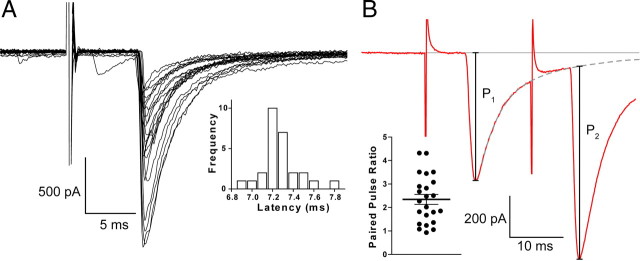
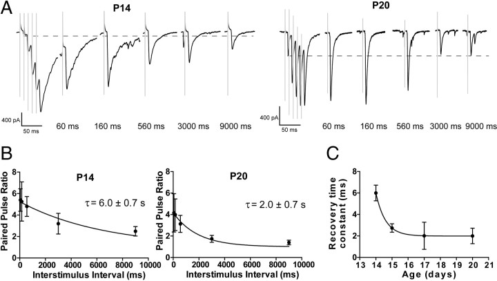
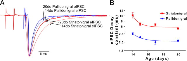
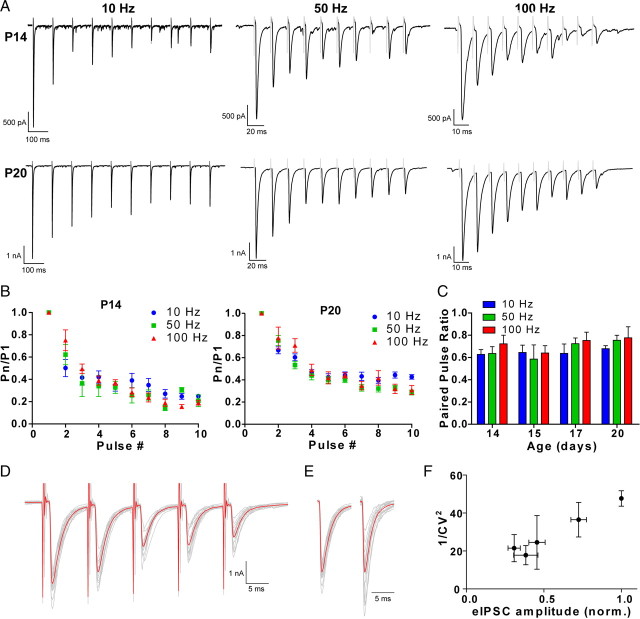
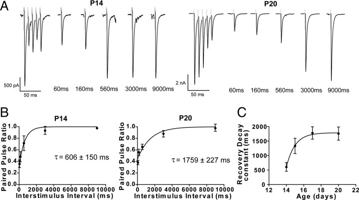
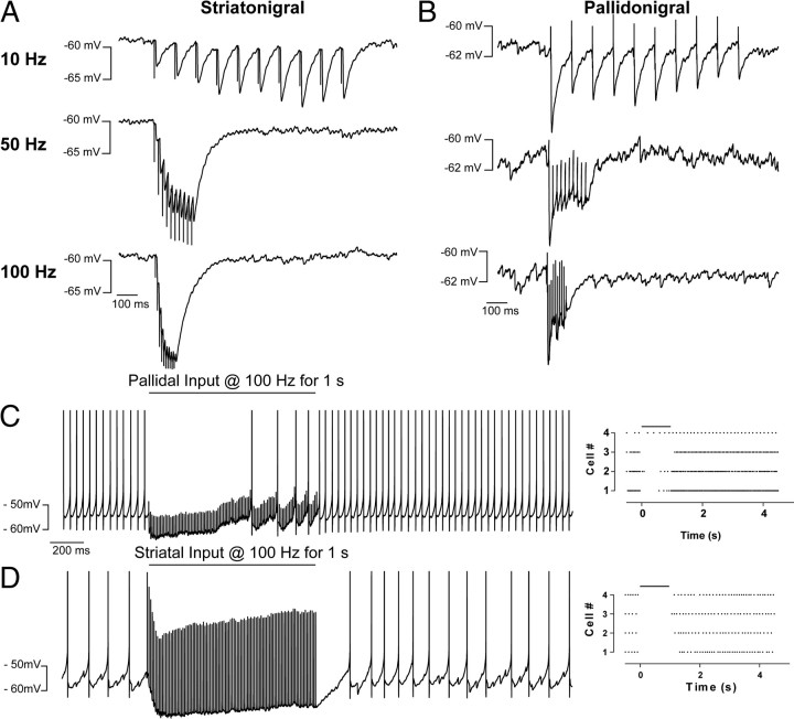
Inhibitory projections from the striatum and globus pallidus converge onto GABAergic projection neurons of the substantia nigra pars reticulata (SNr). Based on existing structural and functional evidence, these pathways are likely to differentially regulate the firing of SNr neurons. We sought to investigate the functional differences in inhibitory striatonigral and pallidonigral traffic using whole-cell voltage clamp in brain slices with these pathways preserved. We found that striatonigral IPSCs exhibited a high degree of paired-pulse facilitation. We tracked this facilitation over development and found the facilitation as the animal aged, but stabilized by postnatal day 17 (P17), with a paired pulse ratio of 2. We also found that the recovery from facilitation accelerated over development, again, reaching a stable phenotype by P17. In contrast, pallidonigral synapses show paired-pulse depression, and this depression could be solely explained by presynaptic changes. The mean paired-pulse ratio of 0.67 did not change over development, but the recovery from depression slowed over development. Pallidonigral IPSCs were significantly faster than striatonigral IPSCs when measured at the soma. Finally, under current clamp, prolonged bursts of striatal IPSPs were able to consistently silence the pacemaker activity of nigral neurons, whereas pallidal inputs depressed, allowing nigral neurons to reinstate firing. These findings highlight the importance of differential dynamics of neurotransmitter release in regulating the circuit behavior of the basal ganglia.


Figures

Figure 1.
Figure 1.
Slice orientation and physiology of SNr neurons. A, Parasagittal slices were taken at 15° from the midline. Dotted line shows the approximate location of the slice. B, Compound photomicrograph showing the position of the electrodes for striatal stimulation (electrode tips visible) and pallidal stimulation. Also shown is the location of the recording electrode in the SNr (patch electrode is faintly visible). Scale bar, 1 mm. C, A typical action potential recorded from the GABAergic neurons of the SNr and the dopaminergic neurons of the SNc. D, Spike width and tonic spiking frequency plotted against each other showing the distinctive firing patterns of two classes of nigral neurons. All neurons with a GABAergic phenotype were recorded randomly from the SNr. Dopaminergic neurons could only be recorded when pipettes were positioned in the SNc.
Figure 2.
Figure 2.
Baseline characteristics of striatonigral eIPSCs. A, Overlay of numerous eIPSCs induced by stimulating in the striatum and recording in the SNr. Inset, Distribution of eIPSC latencies showing their tight distribution. B, Striatonigral IPSCs facilitate. When an eIPSC was evoked during the decaying phase of the previous IPSC, a (bi)exponential was fit to the decay, and the amplitude of the second event was calculated relative to the extrapolated decay of the first. Inset, The paired-pulse ratio (P2/P1) at 50 Hz calculated across all ages.
Figure 3.
Figure 3.
Striatonigral eIPSCs undergo paired-pulse facilitation, which decreases with age. A, B, A train of 10 striatonigral eIPSCs evoked at 10, 50, and 100 Hz recorded from a P14 (A) and a P20 (B) animal. C, Group statistics showing the large degree of facilitation seen in P14 (n = 3–6) animals and how this declines during high-frequency stimulation. P20 (n = 5–6) animals do not show the same degree of facilitation, and this does not decline at high frequencies. D, The paired-pulse ratio between first and second pulse is unaffected by frequency in the 10–100 Hz range, but as animals age, the degree of facilitation decreases (n = 4–7; p = 0.005). Stimulus artifacts are digitally truncated and shaded.
Figure 4.
Figure 4.
The speed of recovery from paired-pulse facilitation increases over development at striatonigral synapses. A, Waveforms showing the rate of recovery in P14 and P20 animals. Dotted line shows the amplitude of the first event. B, The recovery from facilitation in P14 (n = 4) and P20 (n = 6) proceeds in an exponential manner. C, Group statistics show changes in the rate of recovery over development (n = 4–6; p = 0.03).
Figure 5.
Figure 5.
Minimal stimulation used to activate pallidonigral neurons. A, Steadily increasing stimulus intensity recruited two discernable levels of IPSC amplitude. Inset, Typical 2-s-long trace of data showing spontaneous IPSCS with amplitudes >5 nA. B, Relationship between stimulus intensity and eIPSC amplitude from the cell shown in A. C, Occasionally, minimal stimulation in the GP evoked more than a single synaptic event. A fast, presumably pallidal, event can be seen depressing during a train, whereas a second slow, presumably striatal, event can be seen facilitating. Inset, a, The first event in a train in which mixed events were evoked; b, the last event in the train, scaled to the peak amplitude of the first. The first event, subtracted from last, reveals a synaptic event with a long latency, slow rise time, and slow decay time, characteristic of a striatonigral event.
Figure 6.
Figure 6.
Pallidonigral eIPSCs are significantly faster than striatonigral eIPSCs and rate of decay for both increases over development. A, Scaled overlay aligned to response peak showing the difference in kinetics of striatonigral and pallidonigral eIPSC and the changes in kinetics over development. B, Group comparison showing the large difference in the time constant of the decay of striatonigral and pallidonigral eIPSCs (n = 7–9; p < 0.001).
Figure 7.
Figure 7.
Pallidonigral eIPSCs show paired-pulse depression. A, Pallidonigral eIPSCs evoked at 10, 50, and 100 Hz, at P14 and P20 showing clear paired-pulse depression. B, At P14 (n = 5–6) and P20 (n = 7–8), there is no effect of frequency of PPD. C, Group statistics showing the lack of effect of frequency or age on the paired-pulse ratio (n = 4–8; p > 0.3). D, Overlay of a five pulse train at 50 Hz. Calibration: 1 nA, 5 ms. E, The first and last pulse in the train scaled to the same amplitude to show the increase in the coefficient of variation. Calibration: 5 ms. F, 1/CV2 decreases by ∼50%, the same degree as the amplitude of the synaptic events (n = 3), indicating that PPD is solely attributable to a presynaptic mechanism.
Figure 8.
Figure 8.
The recovery from paired-pulse depression at pallidonigral synapses slows over development. A, Example traces showing the recovery from PPD at P14 and P20. B, The recovery from PPD at P14 (n = 4) and P20 (n = 9) can be fit with an exponential. C, The time constant of the recovery process significantly increases over age (n = 4–9; p = 0.02).
Figure 9.
Figure 9.
Striatonigral and pallidonigral input differentially regulate the pacemaker activity of nigral neurons. When SNr neurons are hyperpolarized, striatonigral inputs produce facilitating IPSPs (A) whereas pallidonigral IPSPs depress (B), even when they summate. C, During 1 s, 100 Hz trains, pallidal inputs initially silence nigral neurons, but in three of four cells, nigral neurons begin firing during the train. Left, Peristimulus time histogram, showing the activity of four nigral neurons during the pallidal stimulus (black line). D, The 1 s, 100 Hz striatonigral trains silence nigral activity during the entire duration of the train.

References

    1. Araki M, McGeer PL, McGeer EG. Striatonigral and pallidonigral pathways studied by a combination of retrograde horseradish peroxidase tracing and a pharmacohistochemical method for gamma-aminobutyric acid transaminase. Brain Res. 1985;331:17–24. - PubMed
    1. Barnes TD, Kubota Y, Hu D, Jin DZ, Graybiel AM. Activity of striatal neurons reflects dynamic encoding and recoding of procedural memories. Nature. 2005;437:1158–1161. - PubMed
    1. Beurrier C, Ben-Ari Y, Hammond C. Preservation of the direct and indirect pathways in an in vitro preparation of the mouse basal ganglia. Neuroscience. 2006;140:77–86. - PubMed
    1. Chang HT, Wilson CJ, Kitai ST. Single neostriatal efferent axons in the globus pallidus: a light and electron microscopic study. Science. 1981;213:915–918. - PubMed
    1. Chevalier G, Deniau JM. Disinhibition as a basic process in the expression of striatal functions. Trends Neurosci. 1990;13:277–280. - PubMed

Publication types

MeSH terms

Substances

LinkOut - more resources