

Role of glucocorticoids in tuning hindbrain stress integration
- PMID: 21048149
- PMCID: PMC2997520
- DOI: 10.1523/JNEUROSCI.0522-10.2010
Role of glucocorticoids in tuning hindbrain stress integration
Abstract
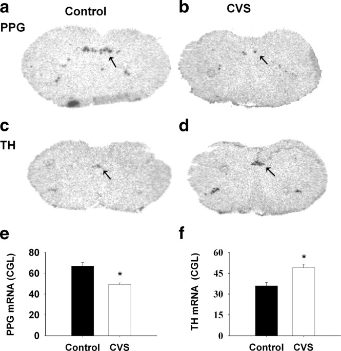
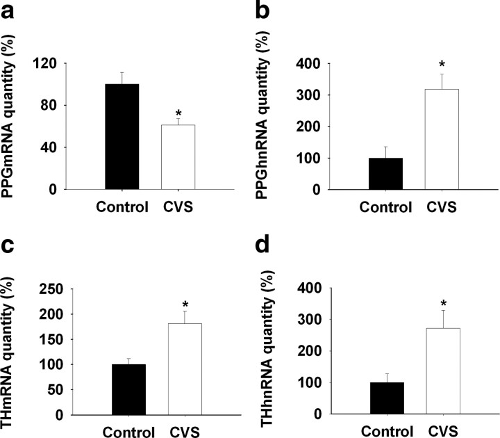
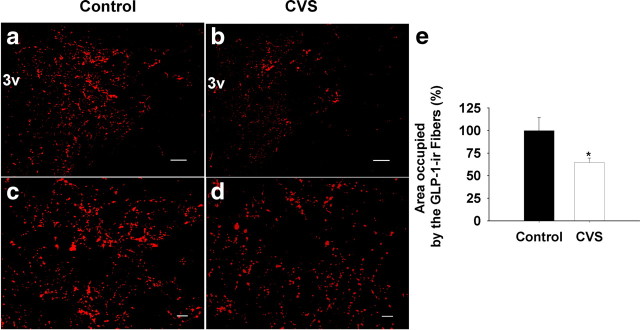
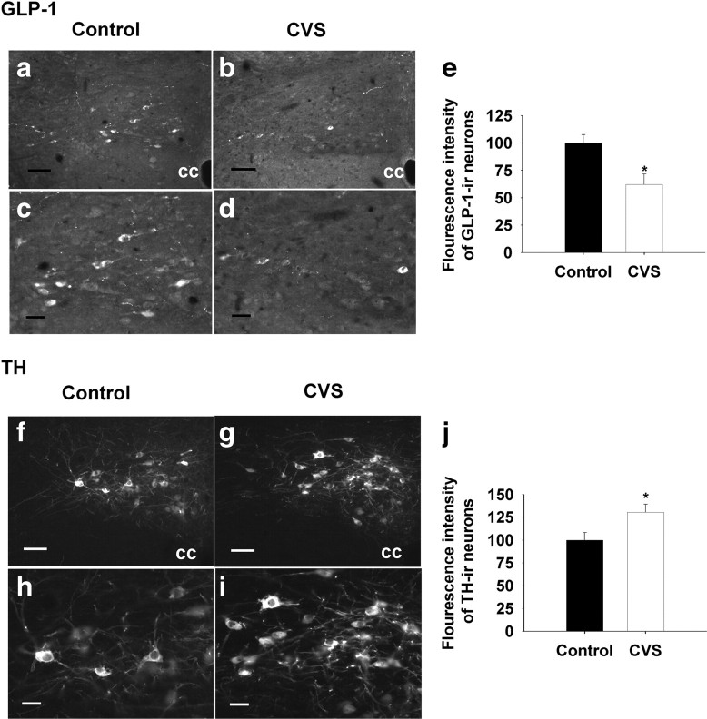
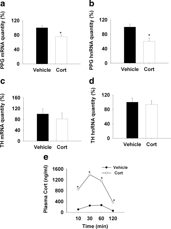
The nucleus of the solitary tract (NTS) is a critical integrative site for coordination of autonomic and endocrine stress responses. Stress-excitatory signals from the NTS are communicated by both catecholaminergic [norepinephrine (NE), epinephrine (E)] and noncatecholaminergic [e.g., glucagon-like peptide-1 (GLP-1)] neurons. Recent studies suggest that outputs of the NE/E and GLP-1 neurons of the NTS are selectively engaged during acute stress. This study was designed to test mechanisms of chronic stress integration in the paraventricular nucleus, focusing on the role of glucocorticoids. Our data indicate that chronic variable stress (CVS) causes downregulation of preproglucagon (GLP-1 precursor) mRNA in the NTS and reduction of GLP-1 innervation to the paraventricular nucleus of the hypothalamus. Glucocorticoids were necessary for preproglucagon (PPG) reduction in CVS animals and were sufficient to lower PPG mRNA in otherwise unstressed animals. The data are consistent with a glucocorticoid-mediated withdrawal of GLP-1 in key stress circuits. In contrast, expression of tyrosine hydroxylase mRNA, the rate-limiting enzyme in catecholamine synthesis, was increased by stress in a glucocorticoid-independent manner. These suggest differential roles of ascending catecholamine and GLP-1 systems in chronic stress, with withdrawal of GLP-1 involved in stress adaptation and enhanced NE/E capacity responsible for facilitation of responses to novel stress experiences.
Figures

Similar articles
-
Activation of PPG neurons following acute stressors differentially involves hindbrain serotonin in male rats.Neuropharmacology. 2021 Apr 1;187:108477. doi: 10.1016/j.neuropharm.2021.108477. Epub 2021 Feb 10. Neuropharmacology. 2021. PMID: 33581143 Free PMC article.
-
Glucocorticoid regulation of preproglucagon transcription and RNA stability during stress.Proc Natl Acad Sci U S A. 2009 Apr 7;106(14):5913-8. doi: 10.1073/pnas.0808716106. Epub 2009 Mar 23. Proc Natl Acad Sci U S A. 2009. PMID: 19307579 Free PMC article.
-
Overnight food deprivation markedly attenuates hindbrain noradrenergic, glucagon-like peptide-1, and hypothalamic neural responses to exogenous cholecystokinin in male rats.Physiol Behav. 2013 Sep 10;121:35-42. doi: 10.1016/j.physbeh.2013.01.012. Epub 2013 Feb 4. Physiol Behav. 2013. PMID: 23391574 Free PMC article.
-
Role of central glucagon-like peptide-1 in stress regulation.Physiol Behav. 2013 Oct 2;122:201-7. doi: 10.1016/j.physbeh.2013.04.003. Epub 2013 Apr 24. Physiol Behav. 2013. PMID: 23623992 Free PMC article. Review.
-
Regulation of Hypothalamo-Pituitary-Adrenocortical Responses to Stressors by the Nucleus of the Solitary Tract/Dorsal Vagal Complex.Cell Mol Neurobiol. 2018 Jan;38(1):25-35. doi: 10.1007/s10571-017-0543-8. Epub 2017 Sep 11. Cell Mol Neurobiol. 2018. PMID: 28895001 Free PMC article. Review.
Cited by
-
Ghrelin signaling contributes to fasting-induced attenuation of hindbrain neural activation and hypophagic responses to systemic cholecystokinin in rats.Am J Physiol Regul Integr Comp Physiol. 2020 May 1;318(5):R1014-R1023. doi: 10.1152/ajpregu.00346.2019. Epub 2020 Apr 15. Am J Physiol Regul Integr Comp Physiol. 2020. PMID: 32292065 Free PMC article.
-
Reporter mouse strain provides a novel look at angiotensin type-2 receptor distribution in the central nervous system.Brain Struct Funct. 2016 Mar;221(2):891-912. doi: 10.1007/s00429-014-0943-1. Epub 2014 Nov 27. Brain Struct Funct. 2016. PMID: 25427952 Free PMC article.
-
Corticosterone Upregulates Gene and Protein Expression of Catecholamine Markers in Organotypic Brainstem Cultures.Int J Mol Sci. 2019 Jun 14;20(12):2901. doi: 10.3390/ijms20122901. Int J Mol Sci. 2019. PMID: 31197099 Free PMC article.
-
BDNF downregulates β-adrenergic receptor-mediated hypotensive mechanisms in the paraventricular nucleus of the hypothalamus.Am J Physiol Heart Circ Physiol. 2019 Dec 1;317(6):H1258-H1271. doi: 10.1152/ajpheart.00478.2019. Epub 2019 Oct 11. Am J Physiol Heart Circ Physiol. 2019. PMID: 31603352 Free PMC article.
-
Neuroendocrine drivers of risk and resilience: The influence of metabolism & mitochondria.Front Neuroendocrinol. 2019 Jul;54:100770. doi: 10.1016/j.yfrne.2019.100770. Epub 2019 Jul 6. Front Neuroendocrinol. 2019. PMID: 31288042 Free PMC article. Review.
References
-
- Ahima RS, Harlan RE. Charting of type II glucocorticoid receptor-like immunoreactivity in the rat central nervous system. Neuroscience. 1990;39:579–604. - PubMed
-
- Akana SF, Cascio CS, Shinsako J, Dallman MF. Corticosterone: narrow range required for normal body and thymus weight and ACTH. Am J Physiol. 1985;249:R527–R532. - PubMed
-
- Akana SF, Dallman MF, Bradbury MJ, Scribner KA, Strack AM, Walker CD. Feedback and facilitation in the adrenocortical system: unmasking facilitation by partial inhibition of the glucocorticoid response to prior stress. Endocrinology. 1992;131:57–68. - PubMed
-
- Barefoot JC, Helms MJ, Mark DB, Blumenthal JA, Califf RM, Haney TL, O'Connor CM, Siegler IC, Williams RB. Depression and long-term mortality risk in patients with coronary artery disease. Am J Cardiol. 1996;78:613–617. - PubMed
-
- Carroll BJ, Curtis GC, Mendels J. Neuroendocrine regulation in depression. II. Discrimination of depressed from nondepressed patients. Arch Gen Psychiatry. 1976;33:1051–1058. - PubMed
Publication types
MeSH terms
Substances
Grants and funding
LinkOut - more resources
Full Text Sources
Medical