. 2010 Nov;177(5):2310-22.
doi: 10.2353/ajpath.2010.100371.

IL-1beta-induced increase in intestinal epithelial tight junction permeability is mediated by MEKK-1 activation of canonical NF-kappaB pathway

Affiliations

IL-1beta-induced increase in intestinal epithelial tight junction permeability is mediated by MEKK-1 activation of canonical NF-kappaB pathway

Rana Al-Sadi et al. Am J Pathol. 2010 Nov.

Abstract

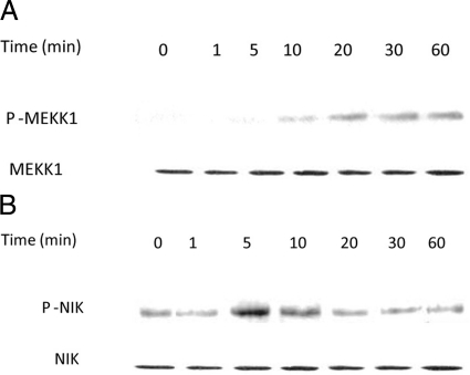
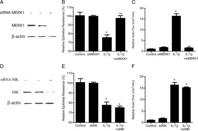
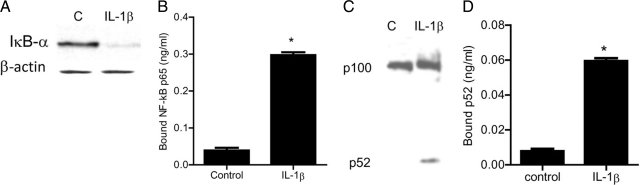
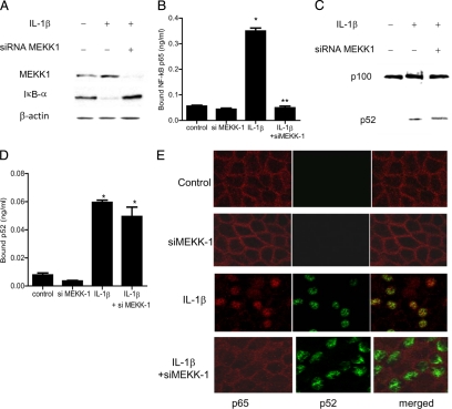
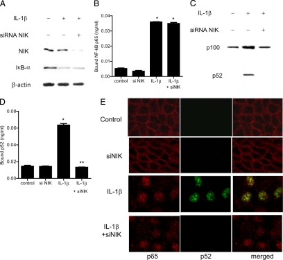
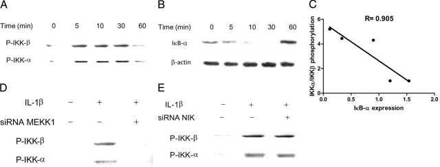
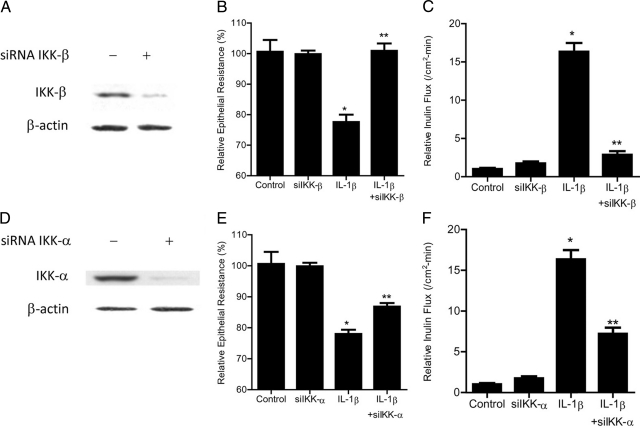
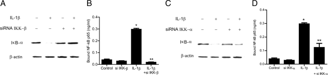
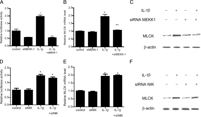
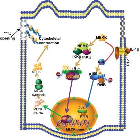
IL-1β is a proinflammatory cytokine that plays a central role in the inflammatory process of the gut. IL-1β causes an increase in intestinal epithelial tight junction (TJ) permeability, but the intracellular pathways that mediate intestinal TJ permeability remain unclear. The major aims of this study were to delineate the protein kinases that regulate the IL-1β modulation of intestinal TJ barrier function and to determine the intracellular mechanisms involved, using filter-grown Caco-2 monolayers as the in vitro model system. Our results showed that IL-1β caused a rapid activation of MEKK-1 and NIK. The knockdown of MEKK-1, but not NIK, inhibited the IL-1β increase in Caco-2 TJ permeability. IL-1β caused an activation of both canonical and noncanonical NF-κB pathways; MEKK-1 regulated the activation of the canonical pathway, while NIK regulated the activation of the noncanonical pathway. Inhibition of MEKK-1 activation of the canonical pathway prevented the IL-1β increase in TJ permeability. Our data also indicated that inhibitory κB kinase was the catalytic subunit primarily involved in canonical pathway activation and TJ barrier opening. MEKK-1 also played an essential role in myosin light chain kinase gene activation. In conclusion, our data show for the first time that MEKK-1 plays an integral role in IL-1β modulation of Caco-2 TJ barrier function by regulating the activation of the canonical NF-κB pathway and the MLCK gene.


Figures

Figure 1
Figure 1
Time course effect of IL-1β on Caco-2 MEKK-1 and NIK activation. A: Time course effect of IL-1β (10 ng/ml) on Caco-2 MEKK-1 phosphorylation (total MEKK-1 was used for equal protein loading). B: Time course effect of IL-1β on NIK phosphorylation (total NIK was used for equal protein loading). IL-1β caused a time-dependent increase in Caco-2 MEKK-1 and NIK activation.
Figure 2
Figure 2
Effect of siRNA-induced MEKK-1 and NIK knockdown of IL-1β–induced increase in Caco-2 TJ permeability. A: MEKK-1 siRNA transfection resulted in a near complete depletion in MEKK-1 protein expression as determined by Western blot analysis. B: MEKK-1 siRNA transfection prevented the IL-1β–induced drop in Caco-2 TER (means ± SE, n = 4). *P < 0.01 versus control; **P < 0.01 versus IL-1β treatment. C: MEKK-1 siRNA transfection prevented the IL-1β–induced increase in inulin flux (means ± SE, n = 4). *P < 0.001 versus control; **P < 0.001 versus IL-1β treatment. D: NIK siRNA transfection resulted in a near complete depletion in NIK protein expression. E: NIK siRNA transfection did not prevent the IL-1β–induced drop in Caco-2 TER (means ± SE, n = 4). *P < 0.01 versus control. F: NIK siRNA transfection did not prevent the IL-1β–induced increase in inulin flux (means ± SE, n = 4). *P < 0.001 versus control.
Figure 3
Figure 3
Effect of IL-1β (10 ng/ml) on Caco-2 NF-κB pathways (p65 and p52) activation. A: IL-1β caused the degradation of IκB-α expression (30-minute experimental period) as assessed by Western blot analysis. B: ELISA-based DNA binding assay of NF-κB p65. IL-1β treatment caused a significant increase in Caco-2-NF-κB p65 binding to the DNA probe. *P < 0.001 versus control. C: IL-1β caused activation of p100 and generation of p52 (30 minutes experimental period) as assessed by Western blot analysis. D: ELISA-based DNA binding assay of NF-κB p52. IL-1β treatment caused a significant increase in Caco-2-NF-κB p52 binding to the DNA probe. *P < 0.001 versus control.
Figure 4
Figure 4
Effect of siRNA-induced MEKK-1 knockdown of IL-1β-activation of NF-κB p65 and p52. A: MEKK-1 siRNA transfection prevented the IL-1β–induced degradation of IκB-α as assessed by Western blot analysis. B: MEKK-1 silencing inhibited the IL-1β–induced binding of p65 to its binding site on the DNA probe as measured by DNA ELISA-binding assay. *P < 0.001 versus control; **P < 0.001 versus IL-1β treatment. C: MEKK-1 siRNA transfection did not prevent the IL-1β generation of p52. D: MEKK-1 silencing did not inhibit the IL-1β–induced binding of p52 to its binding site on DNA probe. *P < 0.001 versus control. E: Effect of MEKK−1 siRNA transfection on IL-1β–induced nuclear translocation of p65 and p52 as viewed by confocal microscopy. Magnification, ×40.
Figure 5
Figure 5
Effect of siRNA-induced NIK knockdown of IL-1β activation of NF-κB p65 and p52. A: NIK siRNA transfection did not prevent the IL-1β–induced degradation of IκB-α as assessed by Western blot analysis. B: NIK silencing did not inhibit the IL-1β–induced binding of p65 to its binding site on DNA probe as measured by DNA ELISA-binding assay. *P < 0.001 versus control. C: NIK siRNA transfection prevented the IL-1β generation of p52. D: NIK silencing inhibited the IL-1β–induced binding of p52 to its binding site on the DNA probe. *P < 0.001 versus control; **P < 0.001 versus IL-1β treatment. E: Effect of NIK siRNA transfection on IL-1β–induced nuclear translocation of p65 and p52 as viewed by confocal microscopy. Magnification, ×40.
Figure 6
Figure 6
Time-course effect of IL-1β on Caco-2 IKK catalytic subunit activation. A: Time-course effect of IL-1β (10 ng/ml) on Caco-2 IKK-α and IKK-β phosphorylation. IL-1β caused a time-dependent increase in Caco-2 IKK-α and IKK-β activation. B: Time-course effect of IL-1β on IκB-α degradation (β-actin was used for equal protein loading). C: Graph of IKK-α/IKK-β activation versus IκB-α degradation (r = 0.905). D: MEKK-1 siRNA transfection prevented the IL-1β–induced phosphorylation of IKK-α and IKK-β as assessed by Western blot analysis. E: NIK siRNA transfection did not prevent the IL-1β–induced phosphorylation of IKK-α and IKK-β.
Figure 7
Figure 7
Effect of siRNA-induced IKK-β or IKK-α knockdown of IL-1β–induced increase in Caco-2 TJ permeability. A: IKK-β siRNA transfection resulted in a near complete depletion in IKK-β protein expression as determined by Western blot analysis. B: IKK-β siRNA transfection completely prevented the IL-1β–induced drop in Caco-2 TER (means ± SE, n = 4). *P < 0.01 versus control; **P < 0.01 versus IL-1β treatment. C: IKK-β siRNA transfection prevented the IL-1β–induced increase in inulin flux (means ± SE, n = 4). *P < 0.001 versus control; **P < 0.001 versus IL-1β treatment. D: IKK-α siRNA transfection resulted in a near complete depletion in IKK-α protein expression as determined by Western blot analysis. E: IKK-α siRNA transfection partially prevented the IL-1β–induced drop in Caco-2 TER. *P < 0.01 versus control; **P < 0.01 versus IL-1β treatment. F: IKK-α siRNA transfection partially prevented the IL-1β–induced increase in inulin flux (means ± SE, n = 4). *P < 0.001 versus control; **P < 0.001 versus IL-1β treatment.
Figure 8
Figure 8
Effect of siRNA IKK subunit knockdown of IL-1β–induced activation of NF-κB p65. A: IKK-β siRNA transfection completely prevented the IL-1β–induced degradation of IκB-α as assessed by Western blot analysis. B: IKK-β siRNA transfection inhibited the IL-1β–induced binding of p65 to its binding site on the DNA probe as measured by DNA ELISA-binding assay. *P < 0.001 versus control; **P < 0.001 versus IL-1β treatment. C: IKK-α siRNA transfection partially prevented the IL-1β–induced degradation of IκB-α as assessed by Western blot analysis. D: IKK-α siRNA transfection partially inhibited the IL-1β–induced binding of p65 to its binding site on the DNA probe as measured by DNA ELISA-binding assay. *P < 0.001 versus control; **P < 0.01 versus IL-1β treatment.
Figure 9
Figure 9
Effect of siRNA induced knockdown of MEKK-1 on IL-1β–induced increase in MLCK gene activity and protein expression. A: MEKK-1 siRNA transfection resulted in a complete inhibition of IL-1β–induced increase in MLCK promoter activity. *P < 0.01 versus control; **P < 0.001 versus IL-1β treatment. B: siRNA MEKK-1 transfection prevented the IL-1β–induced increase in MLCK mRNA levels. *P < 0.01 versus control; **P < 0.01 versus IL-1β treatment. C: MEKK-1 siRNA transfection prevented the IL-1β–induced increase in MLCK protein expression. D: NIK siRNA transfection did not prevent the IL-1β–induced increase in MLCK promoter activity. *P < 0.01 versus control. E: siRNA NIK transfection did not prevent the IL-1β–induced increase in MLCK mRNA levels. *P < 0.01 versus control. F: NIK siRNA transfection did not affect the IL-1β–induced increase in MLCK protein expression.
Figure 10
Figure 10
Proposed scheme of the intracellular pathways involved in IL-1β–induced increase in intestinal epithelial tight junction (TJ) permeability. IL-1β treatment resulted in activation of the MEKK-1 and NIK signaling cascades. MEKK-1 activation resulted in a step-wise activation of IKK and the canonical NF-κB pathway and activation of the MLCK gene, culminating in the opening of the TJ barrier.

References

    1. Hollander D, Vadheim CM, Brettholz E, Petersen GM, Delahunty T, Rotter JI. Increased intestinal permeability in patients with Crohn’s disease and their relatives. A possible etiologic factor. Ann Intern Med. 1986;105:883–885. - PubMed
    1. Ma TY, Anderson JM. Tight junctions and the intestinal barrier. Burlington, MA: Elsevier Academic Press,; Physiology of the Gastrointestinal Tract. 2006:pp1559–1594.
    1. DeMeo MT, Mutlu EA, Keshavarzian A, Tobin MC. Intestinal permeation and gastrointestinal disease. J Clin Gastroenterol. 2002;34:385–396. - PubMed
    1. Hecht G, Koutsouris A, Pothoulakis C, LaMont JT, Madara JL. Clostridium difficile toxin B disrupts the barrier function of T84 monolayers. Gastroenterology. 1992;102:416–423. - PubMed
    1. Turner JR. Intestinal mucosal barrier function in health and disease. Nat Rev Immunol. 2009;9:799–809. - PubMed

Publication types

MeSH terms