. 2010:2010:587213.
doi: 10.4061/2010/587213. Epub 2009 Nov 19.

Acceleration of functional maturation and differentiation of neonatal porcine islet cell monolayers shortly in vitro cocultured with microencapsulated sertoli cells

Affiliations

Acceleration of functional maturation and differentiation of neonatal porcine islet cell monolayers shortly in vitro cocultured with microencapsulated sertoli cells

Francesca Mancuso et al. Stem Cells Int. 2010.

Abstract

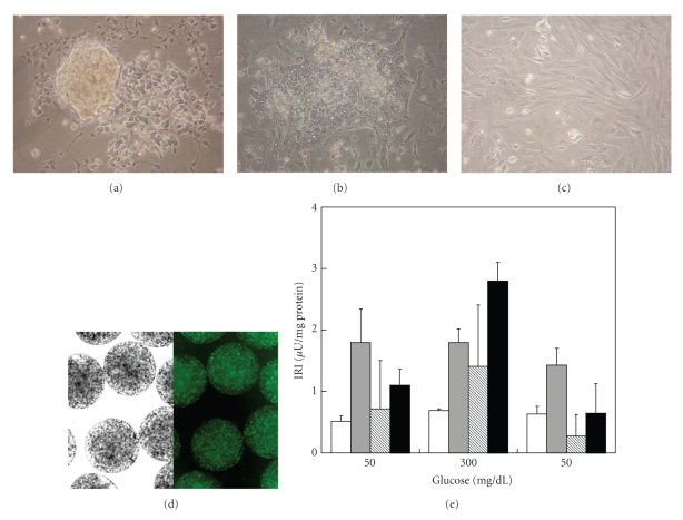
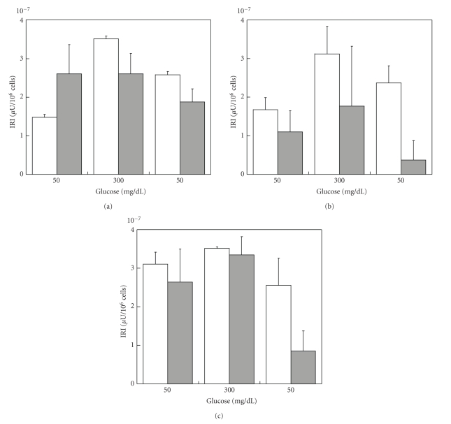
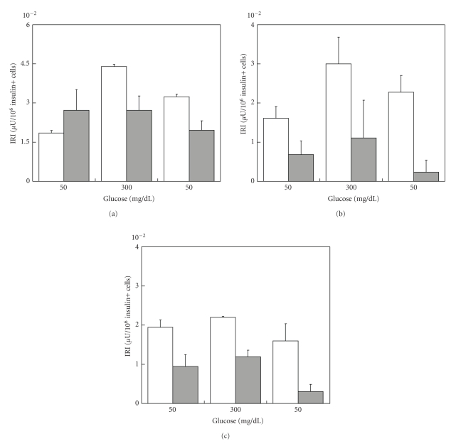
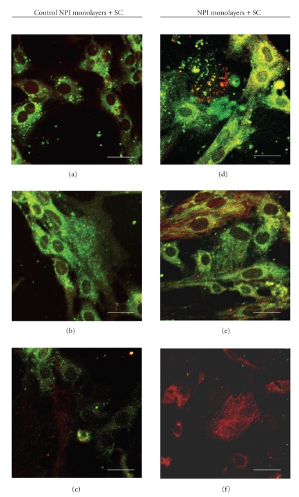
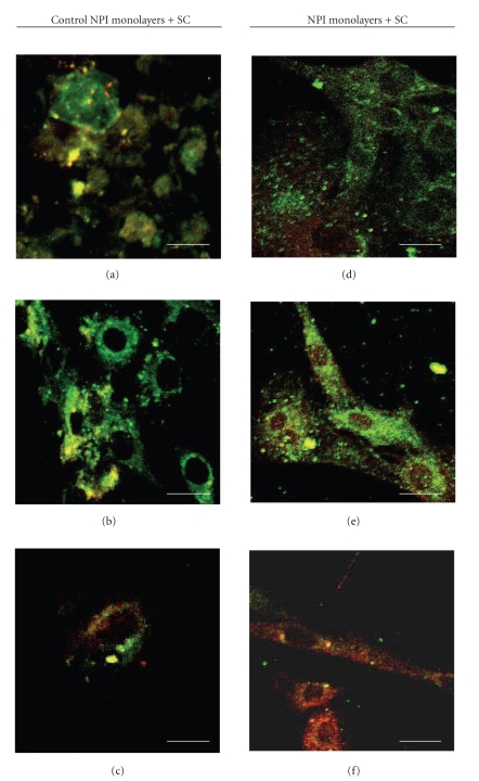
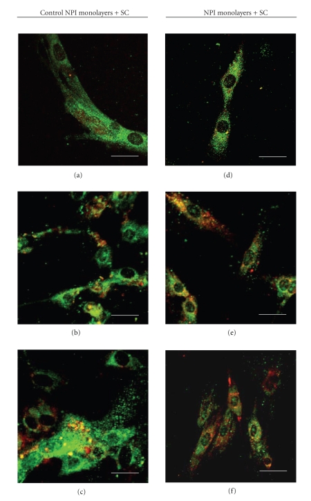
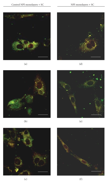
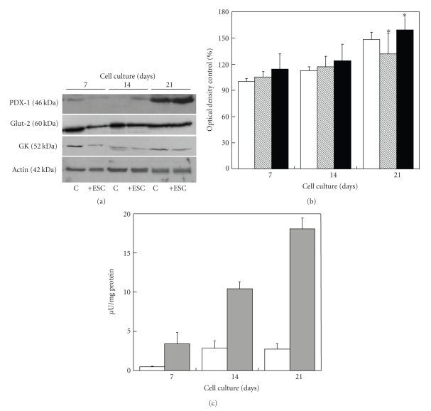
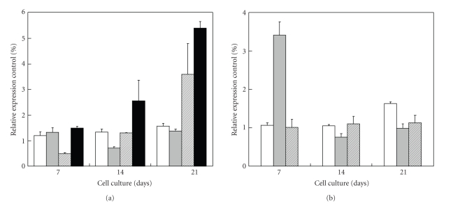
The limited availability of cadaveric human donor pancreata as well as the incomplete success of the Edmonton protocol for human islet allografts fasten search for new sources of insulin the producing cells for substitution cell therapy of insulin-dependent diabetes mellitus (T1DM). Starting from isolated neonatal porcine pancreatic islets (NPIs), we have obtained cell monolayers that were exposed to microencapsulated monolayered Sertoli cells (ESCs) for different time periods (7, 14, 21 days). To assess the development of the cocultured cell monolayers, we have studied either endocrine cell phenotype differentiation markers or c-kit, a hematopoietic stem cell marker, has recently been involved with growth and differentiation of β-cell subpopulations in human as well as rodent animal models. ESC which were found to either accelerate maturation and differentiation of the NPIs β-cell phenotype or identify an islet cell subpopulation that was marked positively for c-kit. The insulin/c-kit positive cells might represent a new, still unknown functionally immature β-cell like element in the porcine pancreas. Acceleration of maturation and differentiation of our NPI cell monolayers might generate a potential new opportunity to develop insulin-producing cells that may suite experimental trials for cell therapy of T1DM.


Figures

Figure 1
Figure 1
(a)–(c) Photomicrographs of NPI after culture for 1 (a), 6 (b), and 10 (c) days on T25 tissue flasks for adherent cell growth. (d) Light field (left) and fluorescence (right) photomicrographs of Ba-AG microcapsules containing SC. Fluorescence micrographs were obtained after staining with EB+FDA to assess SC viability. (e) Insulin secretory patterns of control NPI cell monolayers alone after 14 days (open bars) or NPI cell monolayers, cocultivated with microencapsulated SC for 7 (gray bars), 14 (hatched bars), and 21 days (filled bars) of culture. During static incubation, the cells were treated with the indicated concentrations of glucose. Data represent the average of 3 independent experiments; each insulin determination was performed in triplicate ±SD.
Figure 2
Figure 2
Ratio insulin/total cell number of control NPI cell monolayers alone (open bars), or NPI cell monolayers cocultivated with microencapsulated SC (gray bars) for 7 (a), 14 (b), and 21 days (c). Data are shown as means ± SD from 3 samples.
Figure 3
Figure 3
Ratio insulin/insulin+ cell number of control NPI cell monolayers alone (open bars), or NPI cell monolayers cocultivated with microencapsulated SC (gray bars) for 7 (a), 14 (b), and 21 days (c). Data are shown as means ± SD from 3 samples.
Figure 4
Figure 4
Double fluorescence immunolabeling (green signal: anti-CK-7 Mo-Ab; red signal anti-insulin polyclonal Ab) under confocal laser microscopy of NPI cell monolayers cultivated for 7 (a), (d), 14 (b), (e), and 21 (c), (f) days, alone (a)–(c) or with SC (d)–(f). Bar =10 μm.
Figure 5
Figure 5
Double fluorescence immunolabeling (green signal: anti-PDX-1 Ab; red signal anti-insulin Ab) under confocal laser microscopy of NPI cell monolayers cultivated for 7 (a), (d), 14 (b), (e), and 21 (c), (f) days, alone (a)–(c) or with SC (d)–(f). Bar =10 μm.
Figure 6
Figure 6
Double fluorescence immunolabeling (green signal: anti-PDX-1 Ab; red signal anti-c-kit Ab) under confocal laser microscopy of NPI cell monolayers cultivated for 7 (a), (d), 14 (b), (e), and 21 (c), (f) days, alone (a)–(c) or with SC (d)–(f). Bar =10 μm.
Figure 7
Figure 7
Double fluorescence immunolabeling (green signal: anti-c-kit Ab; red signal anti-insulin Ab) under confocal laser microscopy of NPI cell monolayers cultivated for 7 (a), (d), 14 (b), (e), and 21 (c), (f) days, alone (a)–(c) or with SC (d)–(f). Bar =10 μm.
Figure 8
Figure 8
(a) Western blot analysis of the indicated proteins from control NPI cell monolayers (C) or NPI cell monolayers cocultured with ESC (+ESC) for the indicated time periods. (b) The relative intensities of PDX-1 (open bars), Glut-2 (hatched bars), and GK (filled bars) levels, as determined by band densitometric analysis. The ratio of the band intensities was expressed as a percentage with respect to untreated control NPI cell monolayers. Data are shown as means ± SD from 3 samples. *Significant difference between two groups at each time point (P < .05). (c) Ratio insulin content/mg total protein of control NPI cell monolayers alone (open bars), or NPI cell monolayers, cocultivated with microencapsulated SC (gray bars), for 7, 14, and 21 days of culture. Data are shown as means ± SD from 3 samples.
Figure 9
Figure 9
qPCR analysis of the indicated genes from NPI cell monolayers cocultivated in the presence of ESC for the indicated time periods. (a) PDX-1 (open bars), NeuroD (gray bars), Nkx6.1 (hatched bars), and c-kit (filled bars). (b) Insulin (open bars), Glut-2 (hatched bars), and GK (filled bars). The reported results are expressed as a percentage out of untreated control NPI cell monolayers. Data are shown as means ± SD from 3 samples.

Similar articles

Cited by

References

    1. Nathan DM. The rationale for glucose control in diabetes mellitus. Endocrinology and Metabolism Clinics of North America. 1992;21(2):221–235. - PubMed
    1. Hemkens LG, Grouven U, Bender R, et al. Risk of malignancies in patients with diabetes treated with human insulin or insulin analogues: a cohort study. Diabetologia. 2009;52(9):1732–1744. - PMC - PubMed
    1. Shapiro AMJ, Ricordi C, Hering BJ, et al. International trial of the Edmonton protocol for islet transplantation. The New England Journal of Medicine. 2006;355(13):1318–1330. - PubMed
    1. Korbutt GS, Elliott JF, Ao Z, Smith DK, Warnock GL, Rajotte RV. Large scale isolation, growth, and function of porcine neonatal islet cells. Journal of Clinical Investigation. 1996;97(9):2119–2129. - PMC - PubMed
    1. Luca G, Nastruzzi C, Calvitti M, et al. Accelerated functional maturation of isolated neonatal porcine cell clusters: in vitro and in vivo post-transplant results in NOD mice. Cell Tranplantation. 2005;14:249–261. - PubMed

LinkOut - more resources