. 2010 Oct 15:4:128.
doi: 10.3389/fncel.2010.00128. eCollection 2010.

Enduring medial perforant path short-term synaptic depression at high pressure

Affiliations

Enduring medial perforant path short-term synaptic depression at high pressure

Adolfo E Talpalar et al. Front Cell Neurosci. .

Abstract

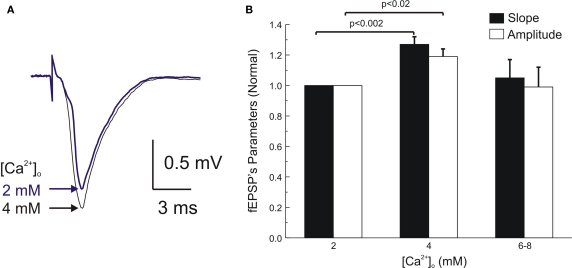
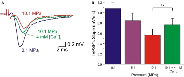
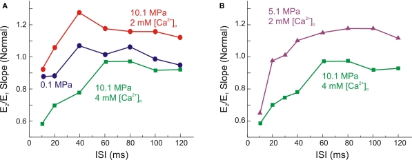
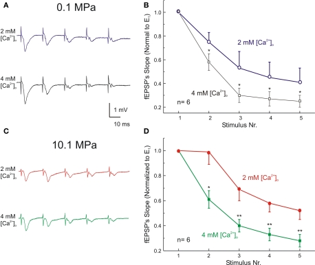
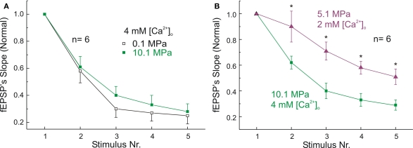
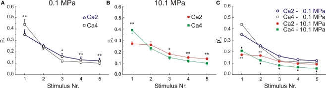
The high pressure neurological syndrome develops during deep-diving (>1.1 MPa) involving impairment of cognitive functions, alteration of synaptic transmission and increased excitability in cortico-hippocampal areas. The medial perforant path (MPP), connecting entorhinal cortex with the hippocampal formation, displays synaptic frequency-dependent-depression (FDD) under normal conditions. Synaptic FDD is essential for specific functions of various neuronal networks. We used rat cortico-hippocampal slices and computer simulations for studying the effects of pressure and its interaction with extracellular Ca(2+) ([Ca(2+)](o)) on FDD at the MPP synapses. At atmospheric pressure, high [Ca(2+)](o) (4-6 mM) saturated single MPP field EPSP (fEPSP) and increased FDD in response to short trains at 50 Hz. High pressure (HP; 10.1 MPa) depressed single fEPSPs by 50%. Increasing [Ca(2+)](o) to 4 mM at HP saturated synaptic response at a subnormal level (only 20% recovery of single fEPSPs), but generated a FDD similar to atmospheric pressure. Mathematical model analysis of the fractions of synaptic resources used by each fEPSP during trains (normalized to their maximum) and the total fraction utilized within a train indicate that HP depresses synaptic activity also by reducing synaptic resources. This data suggest that MPP synapses may be modulated, in addition to depression of single events, by reduction of synaptic resources and then may have the ability to conserve their dynamic properties under different conditions.

Keywords: HPNS; dentate gyrus; entorhinal cortex; granule cells; hippocampus; hyperbaric helium pressure; rat; synaptic dynamics.


Figures

Figure 1
Figure 1
Enhancement of single MPP fEPSP's by elevated [Ca2+]o under normobaric conditions. (A) fEPSPs recorded at the inner dendritic area of the dentate gyrus at [Ca2+]o 2 and 4 mM. (B) Statistical analysis of single fEPSPs amplitudes and slopes under 2, 4, (n = 6) and 6–8 mM [Ca2+]o (n = 4). Note that maximal slope and amplitude were reached at 4 mM [Ca2+]o, apparently reaching saturation since further increase of [Ca2+]o to 6–8 mM resulted in no change or even depression of the fEPSPs (not significantly different from fEPSPs at 2 mM). Scatter bars indicate mean ± SE.
Figure 2
Figure 2
High [Ca2+]o partially antagonizes HP depression of single MPP fEPSP. (A) fEPSPs, recorded at the inner dendritic area of the dentate gyrus, showing the effects of pressure (0.1, 5.1, 10.1 MPa) at 2 mM [Ca2+]o, and the partial restoration of fEPSPs at HP by 4 mM [Ca2+]o. (B) Statistical analysis of the above experiments. fEPSPs are depressed in a pressure-dependent manner. At 10.1 MPa the fEPSP's slope is significantly increased at 4 mM [Ca2+]o (p < 0.0001, n = 8). Further increasing [Ca2+]o to 6 mM paradoxically resulted in reduction of fEPSP's slopes (p < 0.05, n = 2). Scatter bars indicate mean ± SE.
Figure 3
Figure 3
High [Ca2+]o promotes paired-pulse-depression of MPP fEPSPs under HP. (A) Single experiment shows the antagonistic effect of high (4 mM) [Ca2+]o on HP (10.1 MPa) paired-pulse modulation. At 10.1 MPa the initial phase of paired-pulse-depression (PPD) was attenuated, while the later paired-pulse-facilitation (PPF) was increased. Increased [Ca2+]o at HP partially restored the slope of E1, but increased PPD for short ISI and abolished the later phase of PPF for ISI 35–120 ms. (B) Comparison between PPM at 5.1 MPa (2 mM [Ca2+]o) and 10.1 MPa (4 mM [Ca2+]o). Note that E1 under these two conditions is almost equal; however PPD is much greater under the latter condition.
Figure 4
Figure 4
High [Ca2+]o increases frequency-dependent-depression (FDD) at atmospheric pressure. Left: Dendritic recordings showing the effect of [Ca2+]o on trains of 5 fEPSPs (50 Hz) at pressure of 0.1 MPa (A) and 10.1 MPa (C). Calibration bars are the same for both conditions. Right: Statistical analysis of FDD at 50 Hz. Each Ex was normalized with respect to its E1 at its corresponding condition of [Ca2+]o and pressure. Effect of [Ca2+]o on FDD at pressure of 0.1 MPa (B) and at 10.1 MPa (D). *p < 0.05, **p < 0.01 depict statistical difference between stimuli at 2 mM and 4 mM [Ca2+]o (independent t-test). All graphs display means ± SE (n = 6 for each condition).
Figure 5
Figure 5
Effect of pressure on the dynamics of MPP's FDD. (A) Similar FDD is induced by 4 mM [Ca2+]o at pressures of 0.1 MPa and 10.1 MPa. Ex were normalized with respect to their corresponding E1 (n = 6 for each condition; differences at each stimulus were not statistically significant). (B) FDD is more significant at 10.1 MPa – 4 mM [Ca2+]o than at 5.1 MPa – 2 mM [Ca2+]o, (n = 6; *p < 0.05). Note that larger FDD occurs at 10.1 MPa despite of the similarity of E1 under both conditions. All graphs show means ± SE.
Figure 6
Figure 6
Effects of [Ca2+]o on dynamics of MPP synaptic variables during 50 Hz stimulation at two different pressures. (A) High [Ca2+]o significantly increases P1, but it significantly reduces the P3-5 with respect to their respective controls. (B) At 10.1 MPa high [Ca2+]o also increases P1 at expenses of P2–5 denoting reduction of ET. Graphs display means ± SE, n = 6 for each condition *p < 0.05 and **p < 0.01 (paired t-test). (C) Differential effects of [Ca2+]o and pressure on the dynamics of MPP synaptic variables. All the fractions were normalized with respect to ET at control pressure and 4 mM [Ca2+]o, (ET’). The effect of [Ca2+]o and pressure on sequential Ps: P1–2 display significant difference between 0.1 MPa and 10.1 MPa in control [Ca2+]o (n = 6, **p < 0.01). At high [Ca2+]o all Ps were significantly depressed by 10.1 MPa (n = 6, ♦p < 0.05 for all of them). SE bars have been omitted for the sake of clarity.

Similar articles

Cited by

References

    1. Abbott L. F., Varela J. A., Sen K., Nelson S. B. (1997). Synaptic depression and cortical gain control. Science 275, 220–22410.1126/science.275.5297.221 - DOI - PubMed
    1. Abrahamsson T., Gustafsson B., Hanse E. (2005). Synaptic fatigue at the naive perforant path-dentate granule cell synapse in the rat. J. Physiol. 569, 737–75010.1113/jphysiol.2005.097725 - DOI - PMC - PubMed
    1. Abraini J. H. (1997). Inert gas and raised pressure: evidence that motor decrements are due to pressure per se and cognitive decrements due to narcotic action. Pflugers Arch. 433, 788–79110.1007/s004240050346 - DOI - PubMed
    1. Abraini J. H., Ansseau M., Fetchali T. (1993). Pressure-induced disorders in neurotransmission and spontaneous behavior in rats: an animal model of psychosis. Biol. Psychiatr. 34, 622–62910.1016/0006-3223(93)90154-6 - DOI - PubMed
    1. Aravanis A. M., Pyle J. L., Tsien R. W. (2003). Single synaptic vesicles fusing transiently and successively without loss of identity. Nature 423, 643–64710.1038/nature01686 - DOI - PubMed