. 2010 Sep 27:2:130.
doi: 10.3389/fnene.2010.00130. eCollection 2010.

Recent Advances in High-Resolution MR Application and Its Implications for Neurovascular Coupling Research

Affiliations

Recent Advances in High-Resolution MR Application and Its Implications for Neurovascular Coupling Research

Noam Harel et al. Front Neuroenergetics. .

Abstract

The current understanding of fMRI, regarding its vascular origins, is based on numerous assumptions and theoretical modeling, but little experimental validation exists to support or challenge these models. The known functional properties of cerebral vasculature are limited mainly to the large pial surface and the small capillary level vessels. However, a significant lack of knowledge exists regarding the cluster of intermediate-sized vessels, mainly the intracortical, connecting these two groups of vessels and where, arguably, key blood flow regulation takes place. In recent years, advances in MR technology and methodology have enabled the probing of the brain, both structurally and functionally, at resolutions and coverage not previously attainable. Functional MRI has been utilized to map functional units down to the levels of cortical columns and lamina. These capabilities open new possibilities for investigating neurovascular coupling and testing hypotheses regarding fundamental cerebral organization. Here, we summarize recent cutting-edge MR applications for studying neurovascular and functional imaging, both in humans as well as in animal models. In light of the described imaging capabilities, we put forward a theory in which a cortical column, an ensemble of neurons involved in a particular neuronal computation is spatially correlated with a specific vascular unit, i.e., a cluster of an emerging principle vein surrounded by a set of diving arteries. If indeed such a correlation between functional (neuronal) and structural (vascular) units exist as a fundamental intrinsic cortical feature, one could conceivably delineate functional domains in cortical areas that are not known or have not been identified.

Keywords: columns; cortical vessels; fMRI; high-resolution; vascular-unit.


Figures

Figure 1
Figure 1
Maximum intensity projections (MIPs) of the 3D volume of a cat brain viewed in (A) coronal and (B) axial orientations. The axial projection (B) was performed over the full acquisition volume, whereas the coronal projection (A) was produced from a narrow slab indicated by the yellow lines in (B). The coronal view clearly shows the uniform organization of intracortical vessels. The axial view is dominated by the vessels on the pial surface. Figure modified from Bolan et al. (2006).
Figure 2
Figure 2
Vascular model of cat visual cortex. (A) An example of 3D time-of-flight (TOF) microangiography in the cat visual cortex. The axial excitation slab (Slab) was positioned so that its superior edge passed through the sagittal sinus (SS), as can be seen in the coronal view. Arteries, indicated by red arrows, are bright due to unsaturated blood flowing into the slab. Veins, indicated by blue arrows, are dark due to their elevated concentration of deoxyhemoglobin (the BOLD effect). (B) A dorsal view close up showing in greater details vessels classifications using the TOF and BOLD techniques; Veins: blue, Arteries: red. (C) 3D surface rendering of the vessels identified throughout the entire imaged volume. Figure modified from Bolan et al. (2006).
Figure 3
Figure 3
Co-registration of fMRI maps with the underlying vascular model. (A) A representative axial slice across marginal gyrus in the cat primary visual cortex. Orange-to-yellow colors represent voxels that exhibit BOLD signal changes that was correlated with the stimulus paradigm. (B) 3D surface rendering of the veins and arteries (blue and red, respectively) within the imaged volume overlaid with iso-amplitude contour lines of the BOLD signals changes.
Figure 4
Figure 4
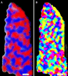
Functional MRI reveals cortical columns in human. An example of ocular dominance (A) and orientation (B) columns fMRI maps obtain in human at 7 T magnet. The red and blue colors in (A) indicate preferences to right or left eye stimulation, whereas the color distribution in (B) represents a given voxel's preferred stimulus orientation. (Scale bar: 1.0 mm) Figure modified from Yacoub et al. (2008).
Figure 5
Figure 5
Whole brain fMRI at high resolution. Functional activation maps for a complex visuomotor dissociation task acquired for 88 slices in 1.25 s with 2 mm × 2 mm × 2 mm resolution. Inset: representative coronal slices from a functional scan with 1 mm × 1 mm × 2 mm resolution. Figure modified from Moeller et al. (2010).
Figure 6
Figure 6
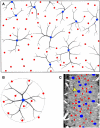
Vascular unit model. A circular nature of the vascular organization. (A) A drawing of a cross-section showing the venous units and their arterial rings. (B) Duvernoy's proposed spatial organization of the vascular unit; a principle cortical vein (blue) surrounded by several penetrating arteries (red). (C) A vessel classification maps in a tangential section in cat visual cortex derived from the MRI data. A and B were modified from Duvernoy et al. (1981).

Similar articles

Cited by

References

    1. Bartfeld E., Grinvald A. (1992). Relationships between orientation-preference pinwheels, cytochrome oxidase blobs, and ocular-dominance columns in primate striate cortex. Proc. Natl. Acad. Sci. U.S.A. 89, 11905–1190910.1073/pnas.89.24.11905 - DOI - PMC - PubMed
    1. Blasdel G. G. (1992). Differential imaging of ocular dominance and orientation selectivity in monkey striate cortex. J. Neurosci. 12, 3115–3138 - PMC - PubMed
    1. Blasdel G. G., Salama G. (1986). Voltage-sensitive dyes reveal a modular organization in monkey striate cortex. Nature 321, 579–58510.1038/321579a0 - DOI - PubMed
    1. Boas D. A., Jones S. R., Devor A., Huppert T. J., Dale A. M. (2008). A vascular anatomical network model of the spatio-temporal response to brain activation. Neuroimage 40, 1116–112910.1016/j.neuroimage.2007.12.061 - DOI - PMC - PubMed
    1. Bolan P. J., Yacoub E., Garwood M., Ugurbil K., Harel N. (2006). In vivo micro-MRI of intracortical neurovasculature. Neuroimage 32, 62–6910.1016/j.neuroimage.2006.03.027 - DOI - PubMed

LinkOut - more resources