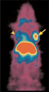
Breast Cancer Treatment in the Era of Molecular Imaging
- PMID: 21048912
- PMCID: PMC2931029
- DOI: 10.1159/000181160
Breast Cancer Treatment in the Era of Molecular Imaging
Abstract
Molecular imaging employs molecularly targeted probes to visualize and often quantify distinct disease-specific markers and pathways. Modalities like intravital confocal or multiphoton microscopy, near-infrared fluorescence combined with endoscopy, surface reflectance imaging, or fluorescence-mediated tomography, and radionuclide imaging with positron emission tomography (PET) and single-photon emission computed tomography (SPECT) are increasingly used for small animal high-throughput screening, drug development and testing, and monitoring gene therapy experiments. In the clinical treatment of breast cancer, PET and SPECT as well as magnetic resonance-based molecular imaging are already established for the staging of distant disease and intrathoracic nodal status, for patient selection regarding receptor-directed treatments, and to gain early information about treatment efficacy. In the near future, reporter gene imaging during gene therapy and further spatial and qualitative characterization of the disease can become clinically possible with radionuclide and optical methods. Ultimately, it may be expected that every level of breast cancer treatment will be affected by molecular imaging, including screening.
Zusammenfassung: Molekulare Bildgebung verwendet auf bestimmte Moleküle gerichtete Sonden, um bestimmte krankheitsspezifische Marker und Stoffwechselwege zu visualisieren und oft auch zu quantifizieren. Modalitäten wie die intravitale konfokale oder Multiphotonenmikroskopie, die Fluoreszenzbildgebung im nahen Infrarot, entweder als oberflächengewichtete Reflexionsbildgebung, als Endoskopie, oder als Fluoreszenz-mediierte Tomographie, und Isotopenbildgebung mit Positronenemissionstomographie (PET) und Single-Photon-Emissionstomographie (SPECT) werden in immer größerem Ausmaß für Kleintiere genutzt. Die Anwendungen umfassen Selektionierungen mit hohem Durchsatz, Medikamentenentwicklung und die Beobachtung von Gentherapieexperimenten. In der klinischen Behandlung von Brustkrebs sind PET und SPECT sowie Magnetresonanz-basierende molekulare Bildgebung bereits für das Staging von Fernmetastasen und für den intrathorakalen Lymphknotentatus etabliert, sowie für die Patientenselektion hinsichtlich Rezeptorgerichteter Therapien und um frühe Informationen über die Effektivität einer Behandlung zu erlangen. In der nahen Zukunft können die Bildgebung von Reportergenen während der Gentherapie und weitere räumliche und qualitative Charakterisierung der Erkrankung mit isotopenbasierten und optischen Methoden klinisch möglich werden. Letztlich kann erwartet werden, dass jede Ebene der Brustkrebsbehandlung von molekularer Bildgebung beeinflusst wird, einschließlich des Screenings.
Figures


Similar articles
-
Recent advances in imaging endogenous or transferred gene expression utilizing radionuclide technologies in living subjects: applications to breast cancer.Breast Cancer Res. 2001;3(1):28-35. doi: 10.1186/bcr267. Epub 2000 Dec 11. Breast Cancer Res. 2001. PMID: 11250742 Free PMC article. Review.
-
Molecular imaging of gene therapy for cancer.Gene Ther. 2004 Aug;11(15):1175-87. doi: 10.1038/sj.gt.3302278. Gene Ther. 2004. PMID: 15141158 Review.
-
Positron Emission Tomography/Magnetic Resonance Imaging for Local Tumor Staging in Patients With Primary Breast Cancer: A Comparison With Positron Emission Tomography/Computed Tomography and Magnetic Resonance Imaging.Invest Radiol. 2015 Aug;50(8):505-13. doi: 10.1097/RLI.0000000000000197. Invest Radiol. 2015. PMID: 26115367
-
Radionuclide-Based Imaging of Breast Cancer: State of the Art.Cancers (Basel). 2021 Oct 30;13(21):5459. doi: 10.3390/cancers13215459. Cancers (Basel). 2021. PMID: 34771622 Free PMC article. Review.
-
Molecular imaging of breast cancer.Breast. 2009 Oct;18 Suppl 3:S66-73. doi: 10.1016/S0960-9776(09)70276-0. Breast. 2009. PMID: 19914546 Review.
References
-
- Weissleder R, Mahmood U. Molecular imaging. Radiology. 2001;219:316–333. - PubMed
-
- Cai W, Gambhir SS, Chen X. Chapter 7. Molecular imaging of tumor vasculature. Methods Enzymol. 2008;445:141–176. - PubMed
-
- Schuster DM, Halkar RK. Molecular imaging in breast cancer. Radiol Clin North Am. 2004;42:885–908. vi-vii. - PubMed
-
- Gambhir SS, Barrio JR, Phelps ME, Iyer M, Namavari M, Satyamurthy N, Wu L, Green LA, Bauer E, MacLaren DC, Nguyen K, Berk AJ, Cherry SR, Herschman HR. Imaging adenoviral-directed reporter gene expression in living animals with positron emission tomography. Proc Natl Acad Sci U S A. 1999;96:2333–2338. - PMC - PubMed
LinkOut - more resources
Full Text Sources

