. 2010 Oct 29;5(10):e13777.
doi: 10.1371/journal.pone.0013777.

Efferent control of the electrical and mechanical properties of hair cells in the bullfrog's sacculus

Affiliations

Efferent control of the electrical and mechanical properties of hair cells in the bullfrog's sacculus

Manuel Castellano-Muñoz et al. PLoS One. .

Abstract

Background: Hair cells in the auditory, vestibular, and lateral-line systems respond to mechanical stimulation and transmit information to afferent nerve fibers. The sensitivity of mechanoelectrical transduction is modulated by the efferent pathway, whose activity usually reduces the responsiveness of hair cells. The basis of this effect remains unknown.

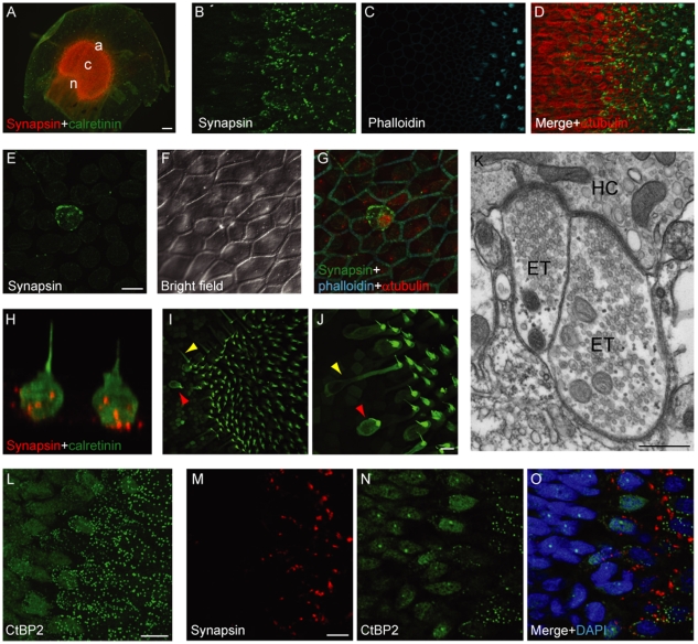
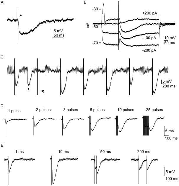
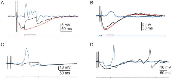
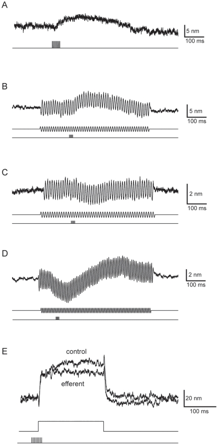
Methodology and principal findings: We employed immunocytological, electrophysiological, and micromechanical approaches to characterize the anatomy of efferent innervation and the effect of efferent activity on the electrical and mechanical properties of hair cells in the bullfrog's sacculus. We found that efferent fibers form extensive synaptic terminals on all macular and extramacular hair cells. Macular hair cells expressing the Ca(2+)-buffering protein calretinin contain half as many synaptic ribbons and are innervated by twice as many efferent terminals as calretinin-negative hair cells. Efferent activity elicits inhibitory postsynaptic potentials in hair cells and thus inhibits their electrical resonance. In hair cells that exhibit spiking activity, efferent stimulation suppresses the generation of action potentials. Finally, efferent activity triggers a displacement of the hair bundle's resting position.

Conclusions and significance: The hair cells of the bullfrog's sacculus receive a rich efferent innervation with the heaviest projection to calretinin-containing cells. Stimulation of efferent axons desensitizes the hair cells and suppresses their spiking activity. Although efferent activation influences mechanoelectrical transduction, the mechanical effects on hair bundles are inconsistent.


Conflict of interest statement

Competing Interests: The authors have declared that no competing interests exist.

Figures

Figure 1
Figure 1. Efferent innervation of saccular hair cells.
A, A wholemount immunofluorescence micrograph shows that synapsin I is highly expressed throughout the saccular macula. The regions used in the statistical analysis are indicated: n, neural; c, central; a, abneural. B D, A higher-magnification stack of confocal images of the macular edge depicts efferent terminals labeled by an anti-synapsin I antiserum (Rb1497) exclusively within the macula, an area demarcated by phalloidin-labeled hair bundles. Note the high expression of alpha-tubulin in extramacular epithelial cells. E G, A maximal-intensity projection of confocal Z-stacks portrays an extramacular hair cell innervated by efferent terminals. H, A three-dimensional confocal reconstruction shows two nearby extramacular hair cells marked by antisera against calretinin (green) and synapsin I (red). I J, A Z-stack projection shows the morphological variability of calretinin-positive hair cells in the macular periphery. Note the occurrence of cylindrical (red arrowheads) and flask-shaped cells (yellow arrowhead). K, As visualized by transmission electron microscopy, two efferent terminals (ET) contact a single hair cell (HC) at adjacent efferent synapses. Note the abundance of synaptic vesicles in the presynaptic terminals and the postsynaptic cisterns in the hair cell. L, A Z-stack projection of the macular periphery shows that an antiserum against CtBP2 labels synaptic ribbons in macular hair cells and nuclei in the epithelial cells outside the macula. M O, A confocal section in the macular periphery shows the abundance of efferent and afferent terminals labeled by respectively an antiserum against synapsin I (Gp118) and one directed against CtBP2. Scale bars: A, 100 µm; D, E, J, L, M, 10 µm; K, 500 nm.
Figure 2
Figure 2. Electrophysiological effect of efferent stimulation.
A, A single shock of the efferent innervation triggered a brief depolarization of a hair cell (arrowhead) followed by long-lasting hyperpolarization (single trace; one compartment, 4 mM Ca2+, resting potential −57 mV). In this and subsequent panels, the stimulus artifact has not been removed. B, The depolarizing component of the response as well as reversed potassium current flow were evident at membrane potentials negative to the equilibrium potential for K+ (single trace; one compartment, 4 mM Ca2+, resting potential −48 mV). C, The electrically evoked inhibitory postsynaptic potentials, which sometimes failed (arrowhead), resembled those obtained spontaneously (asterisk; resting potential −49 mV). D, The magnitude of the inhibitory postsynaptic potential was increased by raising the number of stimulus pulses up to a maximum of five (single traces; one compartment, 4 mM Ca2+). E, The amplitude of the inhibitory postsynaptic potential after pairs of efferent shocks peaked at a separation of 10 ms.
Figure 3
Figure 3. Physiological characteristics of hair cells and electrophysiological effects of efferent stimulation.
A, In response to a depolarizing current pulse (lower trace), electrical resonance (blue trace) was eliminated by a preceding train of efferent stimuli (black trace). The red trace shows the postsynaptic potential in the absence of a current pulse (average of ten presentations, one compartment, 4 mM-Ca2+ saline, resting potential −57 mV). B, In response to a depolarizing current pulse (lower trace), the membrane resistance was reduced by a preceding train of efferent stimuli (black upper trace). The blue trace depicts the control situation with no efferent stimulation; the red trace shows the control situation with no depolarizing current pulse (one compartment, 4 mM-Ca2+ saline, resting potential −53 mV). C, A preceding train of efferent stimulation prevented the occurrence of an action potential in response to a depolarizing current pulse (one compartment, 4 mM-Ca2+ saline, resting potential −57 mV). D, Efferent stimulation blocked rebound spikes and modulated the timing of spontaneous oscillation in the membrane potential (one compartment, 4 mM-Ca2+ saline, resting potential −50 mV).
Figure 4
Figure 4. Effects of efferent stimulation on hair-bundle position.
A, A train of efferent stimuli (lower trace) triggered movement of the hair bundle attached to a compliant probe (upper trace; one compartment, 2 mM-Ca2+ saline). In this and subsequent traces, an upward deflection represents movement in the positive direction, toward the kinocilium. B, During sinusoidal mechanical stimulation (middle traces), the same hair bundle as in A shifted in the positive direction after efferent stimulation without a change in the amplitude of the motion (one compartment, 2 mM-Ca2+ saline, ±30 nm). C, In another hair cell, a similar paradigm induced a shift in the negative direction (two compartments, 0.25 mM-Ca2+ endolymph, 2 mM-Ca2+ saline, ±40 nm). D, Efferent activity occasionally triggered a biphasic movement of the hair bundle (two compartments, 0.25 mM-Ca2+ endolymph, 2 mM-Ca2+ saline, ±40 nm). The traces represent the averages of 20–50 repetitions. E, A hair bundle was stimulated by application of a 150-nm displacement pulse (middle trace) at the base of a compliant fiber. After displacement of the bundle toward its kinocilum, the bundle's movement (top trace) displayed slow adaptation before reaching a steady-state displacement of 44 nm. When the same protocol was preceded by efferent stimulation (bottom trace), the steady-state displacement dropped to 32 nm (two compartments, 0.25 mM-Ca2+ endolymph, 2 mM-Ca2+ saline, fiber stiffness 42 µN m-1, fiber drag coefficient 124 nN s m-1).

Similar articles

Cited by

References

    1. Lewis ER, Baird RA, Leverenz EL, Koyama H. Inner ear: dye injection reveals peripheral origins of specific sensitivities. Science. 1982;215:1641–1643. - PubMed
    1. Rutherford MA, Roberts WM. Spikes and membrane potential oscillations in hair cells generate periodic afferent activity in the frog sacculus. J Neurosci. 2009;29:10025–10037. - PMC - PubMed
    1. Strutz J, Bielenberg K. Efferent acoustic neurons within the lateral superior olivary nucleus of the guinea pig. Brain Res. 1984;299:174–177. - PubMed
    1. Cooper NP, Guinan JJ., Jr Efferent-mediated control of basilar membrane motion. J Physiol. 2006;576:49–54. - PMC - PubMed
    1. Fuchs PA, Parsons TD. The Synaptic Physiology of Hair Cells. In: Eatock RA, Fay RR, Popper AN, editors. Vertebrate Hair Cells. New York: Springer; 2006.

Publication types