BluePort: a platform to study the eosinophilic response of mice to the bite of a vector of Leishmania parasites, Lutzomyia longipalpis sand flies
- PMID: 21048957
- PMCID: PMC2965088
- DOI: 10.1371/journal.pone.0013546
BluePort: a platform to study the eosinophilic response of mice to the bite of a vector of Leishmania parasites, Lutzomyia longipalpis sand flies
Abstract
Background: Visceral Leishmaniasis is a serious human disease transmitted, in the New World, by Lutzomyia longipalpis sand flies. Natural resistance to Leishmania transmission in residents of endemic areas has been attributed to the acquisition of immunity to sand fly salivary proteins. One theoretical way to accelerate the acquisition of this immunity is to increase the density of antigen-presenting cells at the sand fly bite site. Here we describe a novel tissue platform that can be used for this purpose.
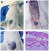
Methodology/principal findings: BluePort is a well-vascularized and macrophage-rich compartment induced in the subcutaneous tissue of mice via injection of agarose beads covered with Cibacron blue. We describe the sequence of inflammatory events leading to its formation and how it can be used to study the dermal response to the bite of L. longipalpis sand flies. Results presented indicate that a shift in the inflammatory response, from neutrophilic to eosinophilic, is the main histopathological feature associated with the immunity acquired through repeated exposure to the bite of sand flies, and that the BluePort tissue compartment could be used to accelerate this process. In addition, changes observed inside the BluePort parenchyma indicate that it could be used to study complex immunobiological processes, and to develop ectopic secondary lymphoid structures.
Conclusions/significance: Understanding the characteristics of the dermal response to the bite of sand flies is a critical element of strategies to control leishmaniasis using vaccines that target salivary proteins. Finding that dermal eosinophilia is such a prominent component of the anti-salivary immunity induced by repeated exposure to sand fly bites raises one important consideration: how to avoid the immunological conflict derived from a protective Th2-driven immunity directed to sand fly saliva with a protective Th1-driven immunity directed to the parasite. The BluePort platform is an ideal tool to address experimentally this conundrum.
Conflict of interest statement
Figures


Similar articles
-
Sand fly salivary proteins induce strong cellular immunity in a natural reservoir of visceral leishmaniasis with adverse consequences for Leishmania.PLoS Pathog. 2009 May;5(5):e1000441. doi: 10.1371/journal.ppat.1000441. Epub 2009 May 22. PLoS Pathog. 2009. PMID: 19461875 Free PMC article.
-
Immunity to a salivary protein of a sand fly vector protects against the fatal outcome of visceral leishmaniasis in a hamster model.Proc Natl Acad Sci U S A. 2008 Jun 3;105(22):7845-50. doi: 10.1073/pnas.0712153105. Epub 2008 May 28. Proc Natl Acad Sci U S A. 2008. PMID: 18509051 Free PMC article.
-
Increased Transmissibility of Leishmania donovani From the Mammalian Host to Vector Sand Flies After Multiple Exposures to Sand Fly Bites.J Infect Dis. 2017 Apr 15;215(8):1285-1293. doi: 10.1093/infdis/jix115. J Infect Dis. 2017. PMID: 28329329 Free PMC article.
-
What's behind a sand fly bite? The profound effect of sand fly saliva on host hemostasis, inflammation and immunity.Infect Genet Evol. 2014 Dec;28:691-703. doi: 10.1016/j.meegid.2014.07.028. Epub 2014 Aug 10. Infect Genet Evol. 2014. PMID: 25117872 Free PMC article. Review.
-
Sand fly saliva: effects on host immune response and Leishmania transmission.Folia Parasitol (Praha). 2006 Sep;53(3):161-71. Folia Parasitol (Praha). 2006. PMID: 17120496 Review.
Cited by
-
Insights into the salivary N-glycome of Lutzomyia longipalpis, vector of visceral leishmaniasis.Sci Rep. 2020 Jul 31;10(1):12903. doi: 10.1038/s41598-020-69753-x. Sci Rep. 2020. PMID: 32737362 Free PMC article.
-
Chemokine-Releasing Microparticles Improve Bacterial Clearance and Survival of Anthrax Spore-Challenged Mice.PLoS One. 2016 Sep 15;11(9):e0163163. doi: 10.1371/journal.pone.0163163. eCollection 2016. PLoS One. 2016. PMID: 27632537 Free PMC article.
References
-
- Sharma U, Singh S. Insect vectors of Leishmania: distribution, physiology and their control. J Vector Borne Dis. 2008;45:255–272. - PubMed
-
- Gramiccia M, Gradoni L. The current status of zoonotic leishmaniases and approaches to disease control. Int J Parasitol. 2005;35:1169–1180. - PubMed
-
- Lainson R, Rangel EF. Lutzomyia longipalpis and the eco-epidemiology of American visceral lesihmaniasis, with particular reference to Brazil: a review. Mem Inst Oswaldo Cruz. 2005;100:811–827. - PubMed
-
- Pearson RD, Sousa AQ. Clinical spectrum of leishmaniasis. Clin Infect Dis. 1996;22:1–13. - PubMed
-
- Reithinger R, Dujardin JC, Luozir H, Pirmez C, Alexander B, et al. Cutaneous lesihmaniasis. Lancet Infect Dis. 2007;7:581–596. - PubMed
Publication types
MeSH terms
Grants and funding
LinkOut - more resources
Full Text Sources
Medical

