. 2010 Oct 27;5(10):e13650.
doi: 10.1371/journal.pone.0013650.

Dexamethasone treatment induces the reprogramming of pancreatic acinar cells to hepatocytes and ductal cells

Affiliations

Dexamethasone treatment induces the reprogramming of pancreatic acinar cells to hepatocytes and ductal cells

Amani Al-Adsani et al. PLoS One. .

Erratum in

Abstract

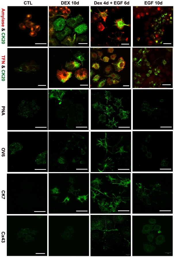
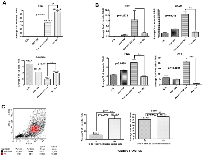
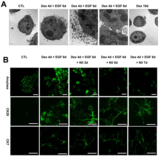
Background: The pancreatic exocrine cell line AR42J-B13 can be reprogrammed to hepatocytes following treatment with dexamethasone. The question arises whether dexamethasone also has the capacity to induce ductal cells as well as hepatocytes.

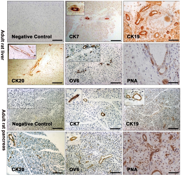
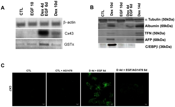
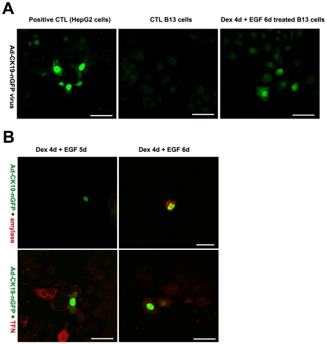
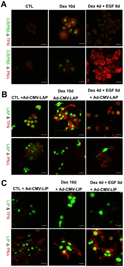
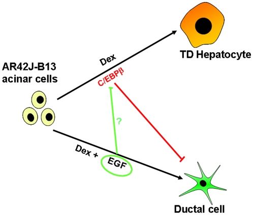
Methodology/principal findings: AR42J-B13 cells were treated with and without dexamethasone and analyzed for the expression of pancreatic exocrine, hepatocyte and ductal markers. Addition of dexamethasone inhibited pancreatic amylase expression, induced expression of the hepatocyte marker transferrin as well as markers typical of ductal cells: cytokeratin 7 and 19 and the lectin peanut agglutinin. However, the number of ductal cells was low compared to hepatocytes. The proportion of ductal cells was enhanced by culture with dexamethasone and epidermal growth factor (EGF). We established several features of the mechanism underlying the transdifferentiation of pancreatic exocrine cells to ductal cells. Using a CK19 promoter reporter, we show that a proportion of the ductal cells arise from differentiated pancreatic exocrine-like cells. We also examined whether C/EBPβ (a transcription factor important in the conversion of pancreatic cells to hepatocytes) could alter the conversion from acinar cells to a ductal phenotype. Overexpression of an activated form of C/EBPβ in dexamethasone/EGF-treated cells provoked the expression of hepatocyte markers and inhibited the expression of ductal markers. Conversely, ectopic expression of a dominant-negative form of C/EBPβ, liver inhibitory protein, inhibited hepatocyte formation in dexamethasone-treated cultures and enhanced the ductal phenotype.

Conclusions/significance: These results indicate that hepatocytes and ductal cells may be induced from pancreatic exocrine AR42J-B13 cells following treatment with dexamethasone. The conversion from pancreatic to hepatocyte or ductal cells is dependent upon the expression of C/EBPβ.


Conflict of interest statement

Competing Interests: The authors have declared that no competing interests exist.

Figures

Figure 1
Figure 1. Expression of ductal markers in adult rat liver and pancreas tissue.
Immunohistochemistry for cytokeratin 7 (CK7), cytokeratin 19 (CK19), cytokeratin 20 (CK20), OV6 and Peanut Agglutinin (PNA) in adult rat liver and pancreas sections. A control (no primary antibody) is also shown. All scale bars, 100 µm.
Figure 2
Figure 2. Differentiation of B13 cells to ductal and hepatic phenotypes.
Immunostaining for Amylase (red)/CK20 (green), TFN (red)/CK20 (green), PNA, OV6, CK7, Cx43 (green) in untreated (control, CTL), DEX, Dex/EGF and EGF treated B13 cells. Scale bars, first and second row, 20 µm; all others 40 µm.
Figure 3
Figure 3. EGF enhances the ductal phenotype at the expense of the hepatic phenotype.
Bar charts showing the percentage of cells expressing (A) Amylase and Transferrin (B) CK7, CK20, PNA and OV-6 in control, EGF, Dex/EGF and Dex treated cells. (C) Scatter plot from the FACSCanto showing the intensity of PNA staining in Dex/EGF treated cells and bar charts showing percentage of cells positive for CK7 and Sox9 (positive fraction) following MACS isolation. Scale bars, top and middle row, 20 µm; lower row, 50 µm.
Figure 4
Figure 4. Expression of ductal markers and inhibition of the ductal phenotype.
(A) RT-PCR for Cx43 and GSTπ (B) Western blotting analysis for Albumin, TFN, AFP and the liver enriched transcription factor C/EBPβ in control, EGF, Dex/EGF and Dex treated cells. β-actin and α-tubulin are also shown as loading controls. (C) Immunostaining for CK7 in control and Dex/EGF treated cells in the presence and absence and absence of the EGF receptor inhibitor AG1478. The inhibitor was added at a final concentration of 25 µM.
Figure 5
Figure 5. Electron microscopy and stability of the ductal phenotype.
(A) Electron micrographs of control, Dex and Dex/EGF treated B13 cells. (B) Immunostaining for amylase, CK20 and CK7 following withdrawal of Dex and EGF in treated B13 cells. Control B13 cells are also shown (B). Scale bars for electron micrographs are (from left to right); 2, 2, 1 and 0.5 µm. Scale bars in second row, 20 µm and 40 µm for all others.
Figure 6
Figure 6. Lineage trace of ductal phenotype.
(A) Infection of HepG2, control B13 and Dex/EGF treated B13 cells with Ad-CK19-nucGFP and (B) immunostaining for amylase and TFN in Dex/EGF treated B13 cells infected with Ad-CK19-nucGFP.
Figure 7
Figure 7. CEBPβ controls the switch in phenotype from pancreatic B13 cells to hepatocyte or ductal cells.
Immunostaining for C/EBPβ/TFN and C/EBPβ/PNA in control, Dex and Dex/EGF treated cells (A) and after infection with Ad-CMV-LAP (B) and Ad-CMV-LIP (C). In A only the induced endogenous C/EBPβ is visible. Scale bars, 20µ m.
Figure 8
Figure 8. Schematic representation of the possible pathways of differentiation of B13 cells into hepatocytes and ductal cells.
Diagram illustrating the potential relationship between pancreatic acinar cells, ductal cells and hepatocytes.

References

    1. Li WC, Yu WY, Quinlan JM, Burke ZD, Tosh D. The molecular basis of transdifferentiation. J Cell Mol Med. 2005;9:569–582. - PMC - PubMed
    1. Reddy JK, Rao MS, Qureshi SA, Reddy MK, Scarpelli DG, et al. Induction and origin of hepatocytes in rat pancreas. J Cell Biol. 1984;98:2082–2090. - PMC - PubMed
    1. Dabeva MD, Hurston E, Shafritz DA. Transcription factor and liver-specific mRNA expression in facultative epithelial progenitor cells of liver and pancreas. Am J Pathol. 1995;147:1633–1648. - PMC - PubMed
    1. Paner GP, Thompson KS, Reyes CV. Hepatoid carcinoma of the pancreas. Cancer. 2000;88:1582–1589. - PubMed
    1. Cingolani N, Shaco-Levy R, Farruggio A, Klimstra DS, Rosai J. Alpha-fetoprotein production by pancreatic tumors exhibiting acinar cell differentiation: study of five cases, one arising in a mediastinal teratoma. Hum Pathol. 2000;31:938–944. - PubMed

Publication types