. 2010 Oct 26;5(10):e13639.
doi: 10.1371/journal.pone.0013639.

Expression of GABAergic receptors in mouse taste receptor cells

Affiliations

Expression of GABAergic receptors in mouse taste receptor cells

Margaret R Starostik et al. PLoS One. .

Abstract

Background: Multiple excitatory neurotransmitters have been identified in the mammalian taste transduction, with few studies focused on inhibitory neurotransmitters. Since the synthetic enzyme glutamate decarboxylase (GAD) for gamma-aminobutyric acid (GABA) is expressed in a subset of mouse taste cells, we hypothesized that other components of the GABA signaling pathway are likely expressed in this system. GABA signaling is initiated by the activation of either ionotropic receptors (GABA(A) and GABA(C)) or metabotropic receptors (GABA(B)) while it is terminated by the re-uptake of GABA through transporters (GATs).

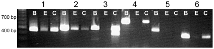
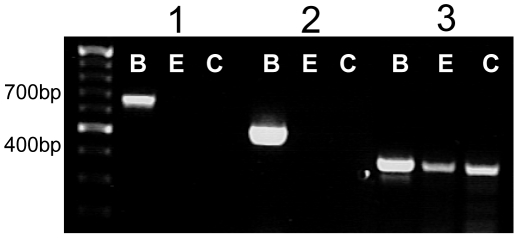
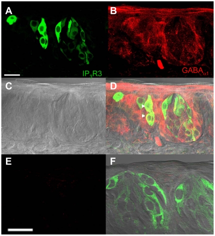
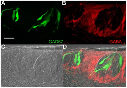
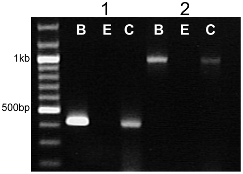
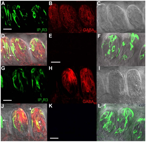
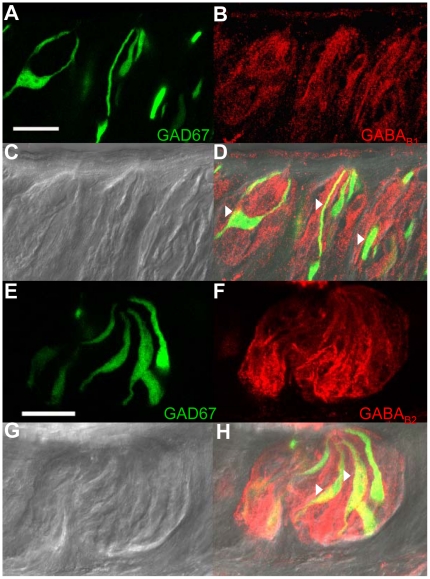
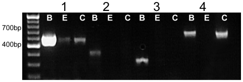
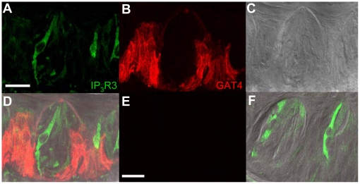
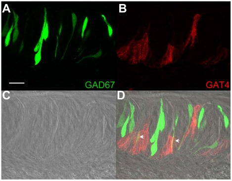
Methodology/principal findings: Using reverse transcriptase-PCR (RT-PCR) analysis, we investigated the expression of different GABA signaling molecules in the mouse taste system. Taste receptor cells (TRCs) in the circumvallate papillae express multiple subunits of the GABA(A) and GABA(B) receptors as well as multiple GATs. Immunocytochemical analyses examined the distribution of the GABA machinery in the circumvallate papillae. Both GABA(A)-and GABA(B)- immunoreactivity were detected in the peripheral taste receptor cells. We also used transgenic mice that express green fluorescent protein (GFP) in either the Type II taste cells, which can respond to bitter, sweet or umami taste stimuli, or in the Type III GAD67 expressing taste cells. Thus, we were able to identify that GABAergic receptors are expressed in some Type II and Type III taste cells. Mouse GAT4 labeling was concentrated in the cells surrounding the taste buds with a few positively labeled TRCs at the margins of the taste buds.

Conclusions/significance: The presence of GABAergic receptors localized on Type II and Type III taste cells suggests that GABA is likely modulating evoked taste responses in the mouse taste bud.


Conflict of interest statement

Competing Interests: The authors have declared that no competing interests exist.

Figures

Figure 1
Figure 1. RT-PCR analysis of the GABAAα subunits.
cDNA from circumvallate TRCs (C), non-gustatory lingual epithelium (E), and brain tissue (B) were subjected to PCR analysis using gene specific primers for the GABAAα subunits 1–6. PCR products were separated by agarose gel electrophoresis. Bands were observed for all subunits in the control brain tissue at the appropriate sizes (α1–455 bp, α2–498 bp, α3–511 bp, α4–699 bp, α5–465 bp, α6-358 bp) while only GABAAα1, GABAAα2, GABAAα3, GABAAα4, and GABAAα6 subunits were detected in taste buds. GABAAα1 and GABAAα2 were detected in the non-gustatory lingual epithelium. Results were repeated at least three times and example data are shown.
Figure 2
Figure 2. RT-PCR analysis of the GABAAβ subunits.
cDNA from circumvallate TRCs (C), non-gustatory lingual epithelium (E), and brain tissue (B) were subjected to PCR analysis using gene specific primers for the GABAA β subunits 1–3. PCR products were separated by agarose gel electrophoresis. Bands were observed for all subunits in the control brain tissue at the appropriate sizes (β1–665 bp, β2–514 bp, β3–415 bp), but only GABAAβ3 was amplified in the TRCs and non-gustatory epithelium. Results were repeated at least three times and representative data are shown.
Figure 3
Figure 3. Localization of GABAAα1 receptors in the IP3R3-GFP expressing circumvallate taste buds.
A Z-stack of 4 laser scanning confocal micrographs (LSCM, 0.5 µm each, collected 1 µm apart) of circumvallate taste buds from an IP3R3-GFP mouse labeled with an antibody directed against the GABAAalpha1 subunit is shown. Panel A shows the GFP fluorescence with the corresponding anti-GABAAα1 immunoreactivity of the same section shown in panel B (red labeling). A DIC bright field image of the taste buds is shown in C. An overlay of the images from A, B, and C is shown in D and demonstrates that some IP3R3-GFP expressing taste cells were immunoreactive for GABAAα1 (see arrowheads for example cells). The lack of labeling when the section is incubated with primary antibody that has been pre-incubated with blocking peptide is shown in E. All staining is eliminated when the blocking peptide is present. F shows an overlay of the panel from E with the corresponding DIC image and GFP expression in the taste buds. Scale bars  = 20 µm.
Figure 4
Figure 4. Localization of GABAAα1 receptors in the GAD67-GFP expressing circumvallate taste buds.
A Z-stack of 5 LSCMs (0.5 µm each, collected 1 µm apart) of circumvallate taste buds from a GAD67-GFP mouse labeled with an antibody directed against the GABAAα1 subunit is shown. Panel A shows the GFP fluorescence with the corresponding anti-GABAAα1 immunoreactivity of the same section shown in panel B (red labeling). A DIC bright field image of the taste buds is shown in C. An overlay of the images from A, B, and C is shown in D. Negative controls are the same as shown in Figure 3. Scale bars  = 20 µm.
Figure 5
Figure 5. RT-PCR analysis of the GABAB subunits.
cDNA from circumvallate TRCs (C), non-gustatory lingual epithelium (E) and brain tissue (B) were subjected to PCR analysis using gene specific primers for the GABAB subunits 1 and 2. Both subunits (B1-427 bp, B2-993 bp) were amplified in the TRCs and brain tissue but not in the lingual epithelium. Results were repeated at least three times and representative data are shown.
Figure 6
Figure 6. Localization of GABAB receptors in the circumvallate taste buds from an IP3R3-GFP expressing mouse.
A Z-stack of 4 LSCMs (0.5 µm each, collected 1 µm apart) of mouse circumvallate taste buds with GFP expression in the IP3R3 expressing taste cells is shown in panel A. The corresponding labeling with anti-GABAB1 is shown in panel B and the DIC image shown in C. D, When images were combined, labeling of taste cells with anti-GABA B1 had some co-expression with IP3R3-GFP expressing taste cells (see arrowheads). Not all IP3R3-GFP expressing taste cells were immunoreactive (see arrow). The lack of secondary labeling in the negative control is shown in E. F, An overlay of E on the DIC image is shown with the corresponding GFP expression. The results from a parallel experiment using anti-GABA B2 are shown in G–J with the appropriate negative controls in K and L. Scale bars  = 20 µm.
Figure 7
Figure 7. Localization of GABAB receptors in the circumvallate taste buds from a GAD67-GFP mouse.
A Z-stack of 4 LSCMs (0.5 µm each, collected 1 µm apart) from mouse circumvallate taste buds with GFP expression in the GAD67-expressing taste cells is shown in panel A. The corresponding labeling with anti-GABAB1 is shown in panel B and the DIC image shown in C. D, When images were combined, many GAD67-GFP expressing TRCs were labeled with anti-GABA B1 (see arrowheads for example cells). Parallel results using anti-GABA B2 are shown in E–H. Negative controls are the same as those shown in Figure 6. Scale bars  = 20 µm.
Figure 8
Figure 8. RT-PCR analysis of the GABA transporters.
cDNA from circumvallate TRCs (C), non-gustatory lingual epithelium (E), and brain tissue (B) were subjected to PCR analysis using gene specific primers for the GAT transporters 1–4. PCR products were amplified for all subunits in the control brain tissue at the appropriate sizes (1–697 bp, 2–438 bp, 3–354 bp, 4–681 bp) while only GAT1 and GAT4 were detected in the taste cells. GAT1 was also detected in the non-gustatory lingual epithelium. Results were repeated at least three times and example data are shown.
Figure 9
Figure 9. Mouse GAT4 immunoreactivity in mouse IP3R3-GFP expressing circumvallate papillae.
A Z-stack of 5 LSCMs (0.5 µm each, collected 1 µm apart) of mouse circumvallate taste buds with GFP expression identifying the IP3R3-expressing taste cells is shown in panel A. The corresponding image of the taste buds labeled with an anti-rat GAT3 (mouse GAT-4) antibody is shown in B with the corresponding DIC image shown in C. D, An overlay of A, B, and C illustrates that most labeling is localized in the surrounding cells near the basolateral portion of the taste bud and in a few cells in basolateral portion of the taste bud. The lack of labeling in the negative control is shown in E. An overlay of E, the corresponding GFP expression and DIC image is shown in F. Scale bars  = 20 µm.
Figure 10
Figure 10. Mouse GAT4 immunoreactivity in circumvallate papillae from GAD67-GFP mice.
A Z-stack of 5 LSCMs (0.5 µm each, collected 1 µm apart) of mouse circumvallate taste buds with GFP expression identifying the GAD67-expressing taste cells is shown in panel A. The corresponding image of the taste buds labeled with an anti-rat GAT3 (mouse GAT4) antibody is shown in B with the DIC image shown in C. D, The overlay of A, B, and C reveals that most GAT3 labeling is localized in the surrounding cells near the basolateral portion of the taste bud. A few GFP expressing taste cells have some overlap with the mouse GAT4 immunoreactivity (see arrowheads). The negative control is the same as Figure 9. Scale bars  = 20 µm.

Similar articles

Cited by

References

    1. Delay RJ, Roper SD. Ultrastructure of taste cells and synapses in the mudpuppy Necturus maculosus. J Comp Neurol. 1988;277:268–280. - PubMed
    1. Roper SD. Signal transduction and information processing in mammalian taste buds. Pflugers Arch. 2007;454:759–776. - PMC - PubMed
    1. Kinnamon JC, Sherman TA, Roper SD. Ultrastructure of mouse vallate taste buds: III. Patterns of synaptic connectivity. J Comp Neurol. 1988;270:1–10, 56-17. - PubMed
    1. Finger TE, Danilova V, Barrows J, Bartel DL, Vigers AJ, et al. ATP signaling is crucial for communication from taste buds to gustatory nerves. Science. 2005;310:1495–1499. - PubMed
    1. Huang YJ, Maruyama Y, Lu KS, Pereira E, Plonsky I, et al. Mouse taste buds use serotonin as a neurotransmitter. J Neurosci. 2005;25:843–847. - PMC - PubMed

Publication types

LinkOut - more resources