. 2010 Oct 26;5(10):e13632.
doi: 10.1371/journal.pone.0013632.

Androgen action via testicular arteriole smooth muscle cells is important for Leydig cell function, vasomotion and testicular fluid dynamics

Affiliations

Androgen action via testicular arteriole smooth muscle cells is important for Leydig cell function, vasomotion and testicular fluid dynamics

Michelle Welsh et al. PLoS One. .

Abstract

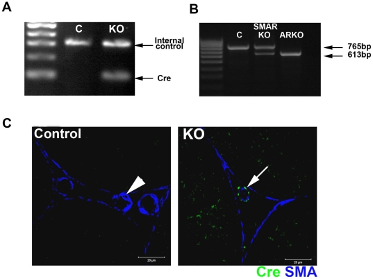
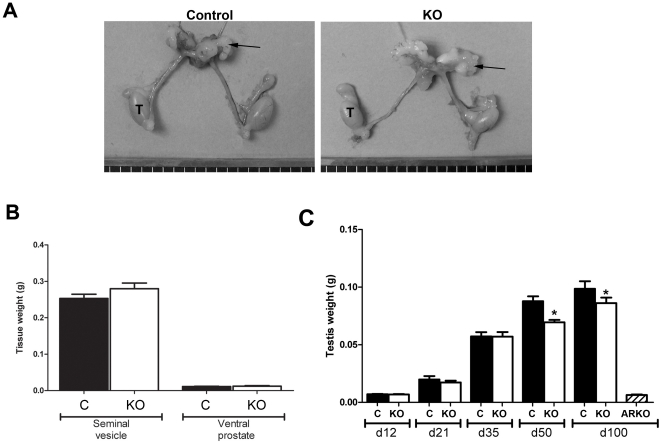
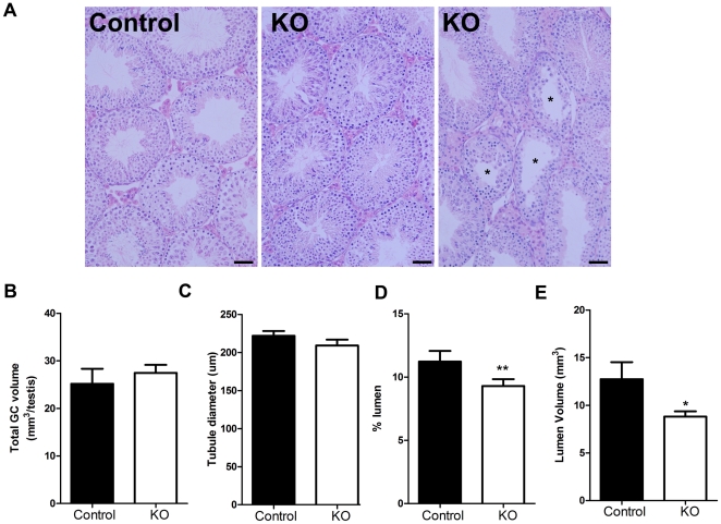
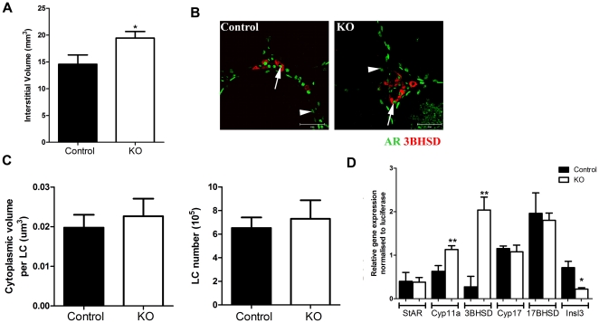
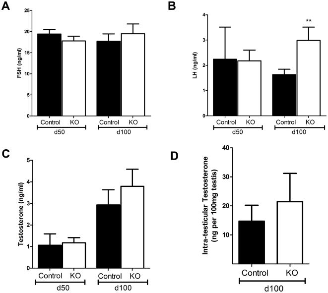
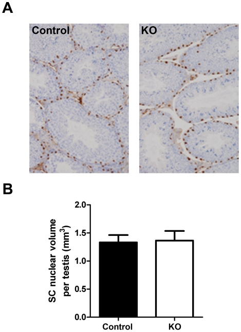
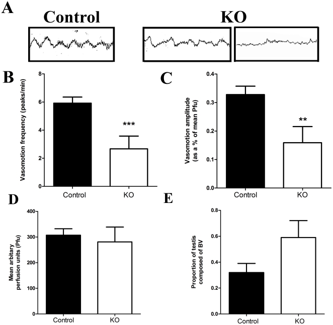
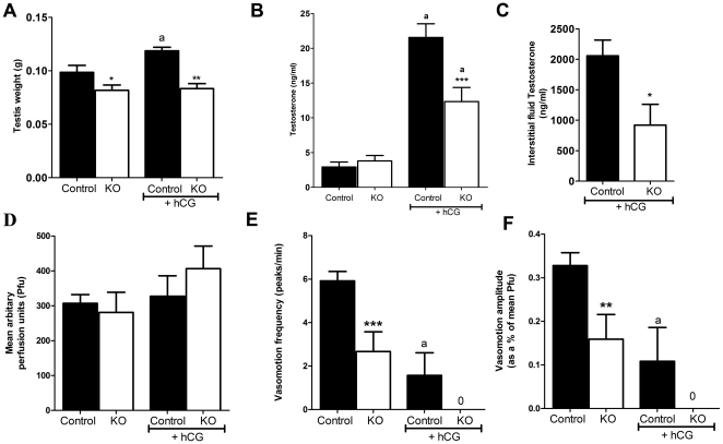
Regulation of blood flow through the testicular microvasculature by vasomotion is thought to be important for normal testis function as it regulates interstitial fluid (IF) dynamics which is an important intra-testicular transport medium. Androgens control vasomotion, but how they exert these effects remains unclear. One possibility is by signalling via androgen receptors (AR) expressed in testicular arteriole smooth muscle cells. To investigate this and determine the overall importance of this mechanism in testis function, we generated a blood vessel smooth muscle cell-specific AR knockout mouse (SMARKO). Gross reproductive development was normal in SMARKO mice but testis weight was reduced in adulthood compared to control littermates; this reduction was not due to any changes in germ cell volume or to deficits in testosterone, LH or FSH concentrations and did not cause infertility. However, seminiferous tubule lumen volume was reduced in adult SMARKO males while interstitial volume was increased, perhaps indicating altered fluid dynamics; this was associated with compensated Leydig cell failure. Vasomotion was impaired in adult SMARKO males, though overall testis blood flow was normal and there was an increase in the overall blood vessel volume per testis in adult SMARKOs. In conclusion, these results indicate that ablating arteriole smooth muscle AR does not grossly alter spermatogenesis or affect male fertility but does subtly impair Leydig cell function and testicular fluid exchange, possibly by locally regulating microvascular blood flow within the testis.


Conflict of interest statement

Competing Interests: The authors have declared that no competing interests exist.

Figures

Figure 1
Figure 1. Characterisation of Cre Recombinase expression in adult SMARKO testes.
A. Approximately 50% of male SMAR were Cre positive (KO), identified by the presence of a band at 100 bp. Control (C) littermates were negative for Cre. B. Deletion of androgen receptor (AR) in the testes was determined using RT-PCR spanning exon 2. Only the larger 765 bp wild-type (WT) AR band was seen in control testes while the smaller 613 bp KO band was seen in ARKO testes. Both bands were identified in SMARKO testes, showing deletion of AR in a proportion of cells. C. Immunohistochemistry for Cre Recombinase (green) and smooth muscle actin (SMA, blue) showed that Cre Recombinase was expressed selectively (arrow) in the smooth muscle cells surrounding the blood vessels in adult SMARKO testes but not in any other testicular cell types or in any cells in control testes (arrowhead).
Figure 2
Figure 2. Gross morphology of the reproductive system from SMARKO mice.
A. Urogenital tract from males at d100 in which the testes (T) are slightly smaller in the SMARKO than in controls while the seminal vesicles (arrow) are not different in size. B. Quantification of seminal vesicle and ventral prostate weight in SMARKO and control (C) mice at d100. C. Quantification of testis weight in SMARKO and control mice from d12-300. Values are means ± S.E.M. (n = 6–22 mice), * p<0.05 compared to controls.
Figure 3
Figure 3. Histological comparison of adult SMARKO and control testes.
A. Most seminiferous tubules in SMARKO testes looked comparable to control testes at d100, however, a small proportion (10%) were abnormal with disturbed spermatogenesis (*). Lumens appeared smaller in many seminiferous tubules in SMARKO testes at d100, compared to controls. B. There was no significant change in the total germ cell volume in SMARKO testes at d100, compared to controls. C. There was no significant change in seminiferous tubule diameter in SMARKO testes at d100, compared to controls. The percentage (D) and volume (E) of seminiferous tubule lumen was significantly reduced at d100 in SMARKO testes compared to controls. Scale bars  = 50 µm. Values are means ± S.E.M. (n = 4–6 mice), * p<0.05, ** p<0.01 compared to controls littermates.
Figure 4
Figure 4. Evaluation of Leydig cell (LC) function in SMARKO testes.
A. There was a significant increase in interstitial volume in SMARKO adult testes. B. Immunostaining for 3βHSD (red) and AR (green) demonstrating that LCs (arrow) and SCs (arrowhead) express AR in both SMARKO and control testes. C. Quantification of LC size and number highlighting no significant change in either in SMARKO testes, compared to controls. D. Relative expression of steroidogenesis enzymes (StAR, cyp11a, 3βHSD, Cyp17 and 17βHSD) and Inls3 in d100 control and SMARKO testes. Values are mean ± SEM (n = 4–6 mice), ** p<0.01, compared to controls littermates. Scale bars  = 50 µm.
Figure 5
Figure 5. Hormone concentrations in SMARKO and control adult males.
A. Serum follicle stimulating hormone (FSH) concentrations were not significantly different in SMARKO males, compared to controls, at d50 or d100. B. Serum luteinising hormone (LH) concentrations were significantly increased in SMARKO males at d100, but not at d50 compared to controls. C. Serum testosterone concentrations were not significantly different in SMARKO males, compared to controls at d50 or d100. D. Intra-testicular testosterone concentrations were not significantly altered in SMARKO males at d100. Values are mean ± SEM (n = 8–27 mice), ** p<0.01, compared to control littermates.
Figure 6
Figure 6. Evaluation of Sertoli cell (SC) function in SMARKO testes.
A. WT-1 staining (black) was used to identify SC nuclei and highlighted their basal location in SMARKO and control males. B. SC mean nuclear volume in SMARKO and control d100 testes. Values are means ± S.E.M. (n = 4–6 mice). Scale bars  = 50 µm.
Figure 7
Figure 7. Vasomotion in SMARKO testes.
A. Representative laser Doppler recordings of testicular blood flow in one control and two KO testes (1 minute each). B. Average vasomotion frequency was significantly reduced in d100 KO testes compared to controls. C. Average vasomotion amplitude was significantly reduced in d100 KO testes compared to controls. D. There was no significant change in average blood flow (Pfu) through d100 SMARKO testes compared to controls. E. Average proportion of blood vessels in d100 control and KO testes. Values are mean ± SEM (n = 3–14 mice), * p<0.05, compared to control littermates.
Figure 8
Figure 8. Testicular response to hCG exposure in SMARKO mice.
A. Testis weight increased significantly in SMAR control, but not in SMARKO 16 hours after hCG treatment, presumably reflecting alterations in interstitial fluid volume. B. Serum testosterone concentrations increased significantly in both SMARKO and control adult males after exposure to hCG, but this increase was larger in controls than in KOs. C. Interstitial fluid testosterone concentrations were significantly higher in control testes than in SMARKOs at d100 after exposure to hCG. D. Average blood flow (Pfu) through adult SMARKO and control testes 16 hours after exposure to hCG. E. Average vasomotion frequency in adult SMARKO and control testes 16 hours after exposure to hCG. F. Average vasomotion amplitude in adult SMARKO and control testes 16 hours after exposure to hCG. Note that vasomotion frequency and amplitude were only measured in the two hcg-treated controls testes in which vasomotion could still be detected. Values are mean ± SEM (n = 5–7 mice), * p<0.05, ** p<0.01, *** p<0.001, compared to control littermates. a p<0.01, compared to untreated mice.

References

    1. George FW, Wilson J. Gonads and ducts in mammals. In: Knobil E, Neill, JD, editors. The Physiology of Reproduction. 2nd ed. New York: Raven Press; 1994. pp. 3–27.
    1. McLachlan RI, O'Donnell L, Meachem SJ, Stanton PG, de Kretser DM, et al. Identification of specific sites of hormonal regulation in spermatogenesis in rats, monkeys, and man. Recent Prog Horm Res. 2002;57:149–179. - PubMed
    1. Sharpe RM. The Physiology of Reproduction. In: Knobil EaN JD, editor. New York: Raven; 1994. pp. 1363–2434.
    1. Quigley CA, De Bellis A, Marschke KB, el-Awady MK, Wilson EM, et al. Androgen receptor defects: historical, clinical, and molecular perspectives. Endocr Rev. 1995;16:271–321. - PubMed
    1. Bremner WJ, Millar MR, Sharpe RM, Saunders PT. Immunohistochemical localization of androgen receptors in the rat testis: evidence for stage-dependent expression and regulation by androgens. Endocrinology. 1994;135:1227–1234. - PubMed

Publication types