

Clinical analysis of high serum IgE in autoimmune pancreatitis
- PMID: 21049558
- PMCID: PMC2975095
- DOI: 10.3748/wjg.v16.i41.5241
Clinical analysis of high serum IgE in autoimmune pancreatitis
Abstract
Aim: To clarify the clinical significance of high serum IgE in autoimmune pancreatitis (AIP).
Methods: Forty-two AIP patients, whose IgE was measured before steroid treatment, were analyzed. To evaluate the relationship between IgE levels and the disease activity of AIP, we examined (1) Frequency of high IgE (> 170 IU/mL) and concomitant allergic diseases requiring treatment; (2) Correlations between IgG, IgG4, and IgE; (3) Relationship between the presence of extrapancreatic lesions and IgE; (4) Relationship between clinical relapse and IgE in patients treated with steroids, and (5) Transition of IgE before and after steroid treatment.
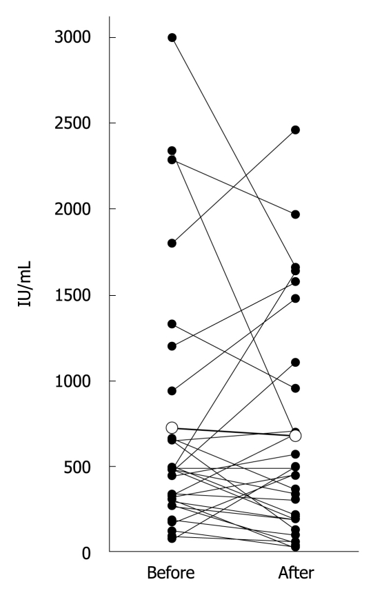
Results: IgE was elevated in 36/42 (86%) patients. Concomitant allergic disease was observed in seven patients (allergic rhinitis in three, bronchial asthma in three, and urticaria in one). There were no significant correlations between IgG, IgG4, and IgE (r = -0.168 for IgG, and r = -0.188 for IgG4). There was no significant difference in IgE in the patients with and without extrapancreatic lesions (526 ± 531 IU/mL vs 819 ± 768 IU/mL, P = 0.163), with and without clinical relapse (457 ± 346 IU/mL vs 784 ± 786 IU/mL, P = 0.374). There was no significant difference in IgE between before and after steroid treatment (723 ± 744 IU/mL vs 673 ± 660 IU/mL, P = 0.633).
Conclusion: Although IgE does not necessarily reflect the disease activity, IgE might be useful for the diagnosis of AIP in an inactive stage.
Figures
References
-
- Shimosegawa T, Kanno A. Autoimmune pancreatitis in Japan: overview and perspective. J Gastroenterol. 2009;44:503–517. - PubMed
-
- Okazaki K, Uchida K, Fukui T. Recent advances in autoimmune pancreatitis: concept, diagnosis, and pathogenesis. J Gastroenterol. 2008;43:409–418. - PubMed
-
- Chari ST, Smyrk TC, Levy MJ, Topazian MD, Takahashi N, Zhang L, Clain JE, Pearson RK, Petersen BT, Vege SS, et al. Diagnosis of autoimmune pancreatitis: the Mayo Clinic experience. Clin Gastroenterol Hepatol. 2006;4:1010–1016; quiz 934. - PubMed
-
- Kawaguchi K, Koike M, Tsuruta K, Okamoto A, Tabata I, Fujita N. Lymphoplasmacytic sclerosing pancreatitis with cholangitis: a variant of primary sclerosing cholangitis extensively involving pancreas. Hum Pathol. 1991;22:387–395. - PubMed
MeSH terms
Substances
LinkOut - more resources
Full Text Sources
Medical
Miscellaneous
