. 2011 Jan 11;50(1):93-105.
doi: 10.1021/bi101288y. Epub 2010 Dec 8.

Structure, energetics, and dynamics of binding coactivator peptide to the human retinoid X receptor α ligand binding domain complex with 9-cis-retinoic acid

Affiliations

Structure, energetics, and dynamics of binding coactivator peptide to the human retinoid X receptor α ligand binding domain complex with 9-cis-retinoic acid

Gang Xia et al. Biochemistry. .

Abstract

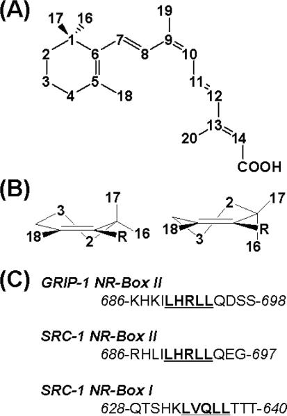
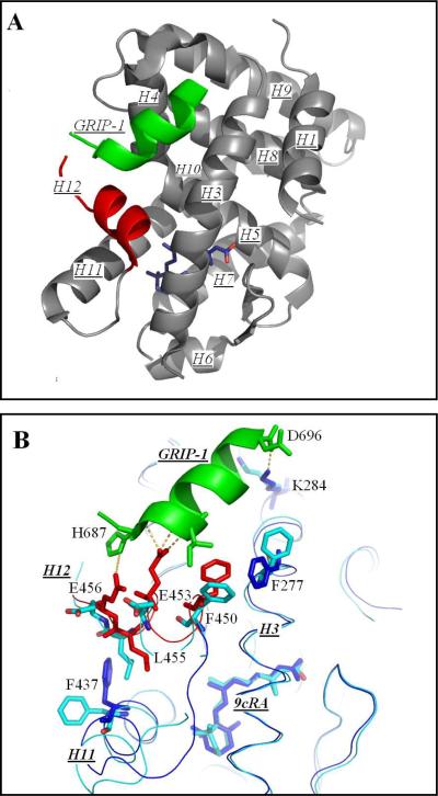
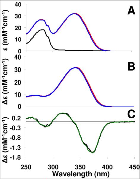
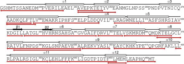
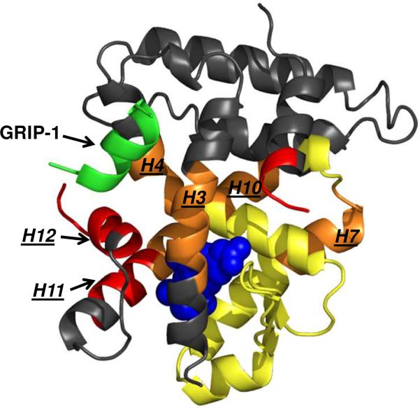
Retinoid X receptors (RXRs) are ligand-dependent nuclear receptors, which are activated by the potent agonist 9-cis-retinoic acid (9cRA). 9cRA binds to the ligand binding domain (LBD) of RXRs and recruits coactivator proteins for gene transcription. Using isothermal titration calorimetry, the binding of a 13-mer coactivator peptide, GRIP-1, to the hRXRα-LBD homodimer complex containing 9cRA (hRXRα-LBD:9cRA:GRIP-1) is reported between 20 and 37 °C. ΔG is temperature independent (-8.5 kcal/mol), and GRIP-1 binding is driven by ΔH (-9.2 kcal/mol) at 25 °C. ΔC(p) is large and negative (-401 cal mol(-1) K(-1)). The crystal structure of hRXRα-LBD:9cRA:GRIP-1 is reported at 2.05 Å. When the structures of hRXRα-LBD:9cRA:GRIP-1 and hRXRα-LBD:9cRA ( 1FBY ) homodimers are compared, E453 and E456 on helix 12 bury and form ionic interactions with GRIP-1. R302 on helix 4 realigns to form new salt bridges to both E453 and E456. F277 (helix 3), F437 (helix 11), and F450 (helix 12) move toward the hydrophobic interior. The changes in the near-UV spectrum at 260 nm of the hRXRα-LBD:9cRA:GRIP-1 support this structural change. Helix 11 tilts toward helix 12 by ≈1 Å, modifying the ring conformation of 9cRA. Hydrogen-deuterium exchange mass spectroscopy indicates GRIP-1 binding to hRXRα-LBD:9cRA significantly decreases the exchange rates for peptides containing helices 3 (F277), 4 (R302), 11 (F437), and 12 (E453, E456). The structural changes and loss of dynamics of the GRIP-1-bound structure are used to interpret the energetics of coactivator peptide binding to the agonist-bound hRXRα-LBD.


Figures

Figure 1
Figure 1
(A) The structure and numbering scheme of 9-cis-retinoic acid. (B) The two half-chair conformations of the trimethylcyclohexenyl ring of 9cRA are shown: the pseudo-axial orientation of the C-17 methyl group in the C-2 down conformation (left), and the pseudo-equatorial orientation of C-17 methyl in the C-2 up conformation (right). (C) The peptide sequences of GRIP-1 NR-Box I and SRC-1 NR-Box I and II coactivator peptides are shown.
Figure 2
Figure 2
Far-UV CD spectrum of hRXRα-LBD:9cRA homodimers in the absence (Solid) and presence (Dashed) of 3-fold excess GRIP-1 coactivator peptide. The far-UV CD spectrum of GRIP-1 coactivator peptide in solution is presented. INSET: The change in mean molar ellipticity at 223 nm for the titration of GRIP-1 into hRXRα-LBD:9cRA homodimers.
Figure 3
Figure 3
The structure of hRXRα-LBD homodimers bound to 9cRA and to GRIP-1. (A) the tertiary conformation of one monomer of the hRXRα-LBD homodimer containing 9cRA (Dark Blue) and coactivator peptide GRIP-1 (Green). Helices 1 through H11 are numbered (H2 missing) and shown in Grey. H12 is colored Red. The amino acid sequences of the helices are: H1 (P231-E243), H3 (D263-R285), H4 (L294-A303), H5 (G304-V320), H6 (R334-S339), H7 (V342-M360), H8 (D363-F376), H9 (N385-K407), H10 (R414-L419), H11 (L422-G443), and H12 (T449-L455). (B) A comparison of hRXRα-LBD:9cRA:GRIP-1 (PDBID-3OAP) and hRXRα-LBD:9cRA (PDBID-1FBY). The structure of hRXRα-LBD:9cRA:GRIP-1 is displayed in Dark Blue for all residues except for H12 which are shown in Red. The structure of hRXRα-LBD:9cRA is displayed in light blue. Significant changes occur for F450, E453, and E456 on H12 (Red), and for F437 on H11 and F277 on H3 (Dark Blue). Yellow dashes refer to new ionic or hydrogen bonding interactions. (C) The structural changes of R302 on H4 (Dark Blue) and E453 and E456 on H12 (Red). New ionic interactions/hydrogen bonds are shown in Yellow. (D) The structural change of the trimethylcyclohexenyl ring of 9cRA in hRXRα-LBD:9cRA:GRIP-1 (Dark Blue) and hRXRα-LBD:9cRA (Light Blue). The trimethylcyclohexenyl ring inverts from one half-chair conformation to the other. Hydrophobic interactions with H11 residues are shown in Dark Blue dashes for the GRIP-1 bound structure and in Light Blue dashes for the structure without GRIP-1.
Figure 3
Figure 3
The structure of hRXRα-LBD homodimers bound to 9cRA and to GRIP-1. (A) the tertiary conformation of one monomer of the hRXRα-LBD homodimer containing 9cRA (Dark Blue) and coactivator peptide GRIP-1 (Green). Helices 1 through H11 are numbered (H2 missing) and shown in Grey. H12 is colored Red. The amino acid sequences of the helices are: H1 (P231-E243), H3 (D263-R285), H4 (L294-A303), H5 (G304-V320), H6 (R334-S339), H7 (V342-M360), H8 (D363-F376), H9 (N385-K407), H10 (R414-L419), H11 (L422-G443), and H12 (T449-L455). (B) A comparison of hRXRα-LBD:9cRA:GRIP-1 (PDBID-3OAP) and hRXRα-LBD:9cRA (PDBID-1FBY). The structure of hRXRα-LBD:9cRA:GRIP-1 is displayed in Dark Blue for all residues except for H12 which are shown in Red. The structure of hRXRα-LBD:9cRA is displayed in light blue. Significant changes occur for F450, E453, and E456 on H12 (Red), and for F437 on H11 and F277 on H3 (Dark Blue). Yellow dashes refer to new ionic or hydrogen bonding interactions. (C) The structural changes of R302 on H4 (Dark Blue) and E453 and E456 on H12 (Red). New ionic interactions/hydrogen bonds are shown in Yellow. (D) The structural change of the trimethylcyclohexenyl ring of 9cRA in hRXRα-LBD:9cRA:GRIP-1 (Dark Blue) and hRXRα-LBD:9cRA (Light Blue). The trimethylcyclohexenyl ring inverts from one half-chair conformation to the other. Hydrophobic interactions with H11 residues are shown in Dark Blue dashes for the GRIP-1 bound structure and in Light Blue dashes for the structure without GRIP-1.
Figure 4
Figure 4
(A) Near UV-Vis spectrum of apo-hRXRα-LBD homodimer (black), hRXRα-LBD:9cRA complex in the absence of GRIP-1 (Red), and hRXRα-LBD:9cRA:GRIP-1 complex (Blue). (B) Difference UV-Vis spectrum of hRXRα-LBD:9cRA complex (Red) or the hRXRα-LBD:9cRA:GRIP-1 complex (Blue) minus the spectrum of apo-hRXRα-LBD. (C). Difference UV-Vis spectrum of hRXRα-LBD:9cRA:GRIP-1 minus the spectrum of hRXRα-LBD:9cRA (Black) versus the computer-fitted difference spectrum (Green).
Figure 5
Figure 5
HDX MS peptides. The 24 HDX MS peptides in Table 3 are mapped onto the hRXRα-LBD protein sequence. Structural features of the hRXRα-LBD are shown above the sequence. Arrows below the sequence correspond to the observed HDX MS peptides in Table 3. An additional 14 overlapping peptides that were observed are provided in Supplemental Table S4.
Figure 6
Figure 6
HDX MS analyses mapped onto hRXRα-LBD:9cRA:GRIP-1. Regions within the hRXRα-LBD that do not change deuterium incorporation with 9cRA or 9cRA + GRIP-1 are colored in grey. Upon 9cRA binding, several HDX MS monitored peptides (yellow and orange) around the ligand binding pocket of hRXRα-LBD become significantly more protected from deuterium incorporation (10–37% change relative to hRXRα-LBD, see Table 3). When GRIP-1 (green) is added to the complex, a subset of these same regions (in orange, H3, H4, H10, and H7) become further protected or stabilized (>7% additional change relative to hRXRα-LBD:9cRA) demonstrating the stabilizing affect of GRIP-1 binding on the hRXRα-LBD beyond the coactivator binding site. Signficantly, several peptides that are part of H12 and the C-terminal end of H11 (in red) were not protected (remained dynamic) by 9cRA addition but then did show protection when GRIP-1 is added to the hRXRα-LBD:9cRA complex.

References

    1. Mangelsdorf DJ, Evans RM. The RXR heterodimers and orphan receptors. Cell. 1995;83:841–850. - PubMed
    1. Tsai MJ, O'Malley BW. Molecular mechanisms of action of steroid/thyroid receptor superfamily members. Annu Rev Biochem. 1994;63:451–486. - PubMed
    1. Mangelsdorf DJ, Thummel C, Beato M, Herrlich P, Schutz G, Umesono K, Blumberg B, Kastner P, Mark M, Chambon P, Evans RM. The nuclear receptor superfamily: the second decade. Cell. 1995;83:835–839. - PMC - PubMed
    1. Dong D, Noy N. Heterodimer formation by retinoid X receptor: regulation by ligands and by the receptor's self-association properties. Biochemistry. 1998;37:10691–10700. - PubMed
    1. Svensson S, Ostberg T, Jacobsson M, Norstrom C, Stefansson K, Hallen D, Johansson IC, Zachrisson K, Ogg D, Jendeberg L. Crystal structure of the heterodimeric complex of LXRalpha and RXRbeta ligand-binding domains in a fully agonistic conformation. Embo J. 2003;22:4625–46233. - PMC - PubMed

Publication types

Associated data

LinkOut - more resources