

Virtual ligand screening of the National Cancer Institute (NCI) compound library leads to the allosteric inhibitory scaffolds of the West Nile Virus NS3 proteinase
- PMID: 21050032
- PMCID: PMC3033206
- DOI: 10.1089/adt.2010.0309
Virtual ligand screening of the National Cancer Institute (NCI) compound library leads to the allosteric inhibitory scaffolds of the West Nile Virus NS3 proteinase
Abstract
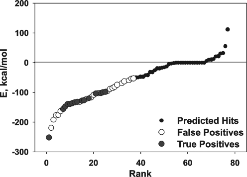
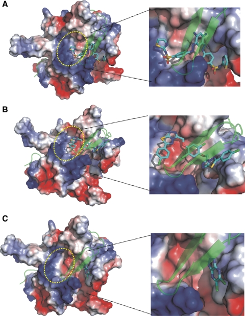
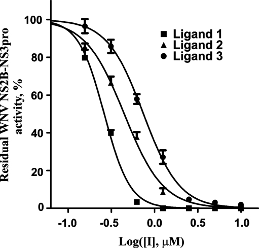
Viruses of the genus Flavivirus are responsible for significant human disease and mortality. The N-terminal domain of the flaviviral nonstructural (NS)3 protein codes for the serine, chymotrypsin-fold proteinase (NS3pro). The presence of the nonstructural (NS)2B cofactor, which is encoded by the upstream gene in the flaviviral genome, is necessary for NS3pro to exhibit its proteolytic activity. The two-component NS2B-NS3pro functional activity is essential for the viral polyprotein processing and replication. Both the structure and the function of NS2B-NS3pro are conserved in the Flavivirus family. Because of its essential function in the posttranslational processing of the viral polyprotein precursor, NS2B-NS3pro is a promising target for anti-flavivirus drugs. To identify selective inhibitors with the reduced cross-reactivity and off-target effects, we focused our strategy on the allosteric inhibitors capable of targeting the NS2B-NS3pro interface rather than the NS3pro active site. Using virtual ligand screening of the diverse, ∼275,000-compound library and the catalytic domain of the two-component West Nile virus (WNV) NS2B-NS3pro as a receptor, we identified a limited subset of the novel inhibitory scaffolds. Several of the discovered compounds performed as allosteric inhibitors and exhibited a nanomolar range potency in the in vitro cleavage assays. The inhibitors were also potent in cell-based assays employing the sub-genomic, luciferase-tagged WNV and Dengue viral replicons. The selectivity of the inhibitors was confirmed using the in vitro cleavage assays with furin, a human serine proteinase, the substrate preferences of which are similar to those of WNV NS2B-NS3pro. Conceptually, the similar in silico drug discovery strategy may be readily employed for the identification of inhibitors of other flaviviruses.
Figures
References
-
- Hanley KA. Weaver SC. Caister Academic Press; Norfolk, UK: 2010. Frontiers in Dengue Virus Research.
-
- Erbel P. Schiering N. D'Arcy A, et al. Structural basis for the activation of flaviviral NS3 proteases from dengue, West Nile virus. Nat Struct Mol Biol. 2006;13:372–373. - PubMed
-
- Chambers TJ. Droll DA. Tang Y, et al. Yellow fever virus NS2B-NS3 protease: characterization of charged-to-alanine mutant and revertant viruses and analysis of polyprotein-cleavage activities. J Gen Virol. 2005;86:1403–1413. - PubMed
-
- Chappell KJ. Stoermer MJ. Fairlie DP. Young PR. West Nile Virus NS2B/NS3 protease as an antiviral target. Curr Med Chem. 2008;15:2771–2784. - PubMed
Publication types
MeSH terms
Substances
Grants and funding
LinkOut - more resources
Full Text Sources
Miscellaneous
