

Vertebrobasilar Flow Evaluation and Risk of Transient Ischaemic Attack and Stroke study (VERiTAS): rationale and design
- PMID: 21050408
- PMCID: PMC3057649
- DOI: 10.1111/j.1747-4949.2010.00528.x
Vertebrobasilar Flow Evaluation and Risk of Transient Ischaemic Attack and Stroke study (VERiTAS): rationale and design
Abstract
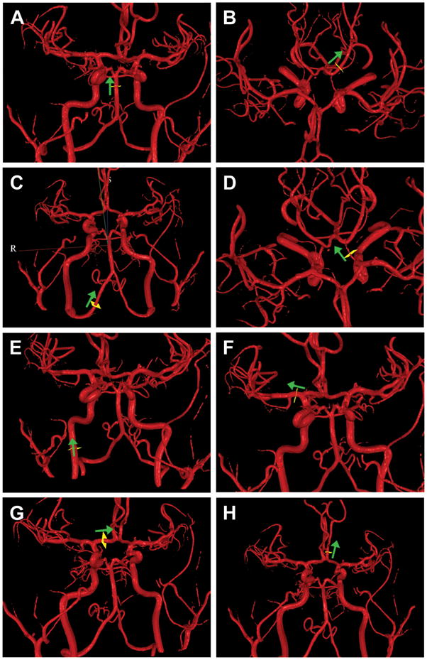
Background: Over one-third of ischaemic strokes occur in the posterior circulation, and a leading cause is atherosclerotic vertebrobasilar disease. Symptomatic vertebrobasilar disease carries a high annual recurrent stroke risk, averaging 10-15% per year. Endovascular angioplasty and stenting are increasingly used but carry risks, and the benefit remains unproven. Determining stroke predictors in this population is critical to identifying high-risk patients for future trials of intervention. Preliminary studies indicate that stroke risk in vertebrobasilar disease is strongly related to haemodynamic compromise, which can be measured noninvasively using quantitative magnetic resonance angiography.
Methods/study design: The Vertebrobasilar Flow Evaluation and Risk of Transient Ischaemic Attack and Stroke (VERiTAS) study, a prospective multicentre NIH-funded observational study of symptomatic vertebrobasilar stenosis (≥50%) or occlusion, is designed to test the hypothesis that patients demonstrating compromised blood flow as assessed by quantitative magnetic resonance angiography are at higher stroke risk. The study will recruit 80 patients at six sites in North America over 4-years. Upon enrollment, subjects will undergo haemodynamic assessment with blinded quantitative magnetic resonance angiography to assess large vessel flow in the vertebrobasilar territory, and be prospectively designated as compromised or normal flow. Patients will be re-imaged with quantitative magnetic resonance angiography at 6-, 12-, and 24-months, and followed for 12-24-months for the primary end-point of stroke in the vertebrobasilar territory.
Conclusion: The VERiTAS study is the first prospective study of haemodynamics and stroke risk in the posterior circulation. The results may impact the selection criteria for interventional candidates and also define a low-risk population in whom the risks of invasive interventions would be unnecessary.
© 2010 The Authors. International Journal of Stroke © 2010 World Stroke Organization.
Figures
References
-
- Whisnant JP, Cartlidge NE, Elveback LR. Carotid and vertebral-basilar transient ischemic attacks: effect of anticoagulants, hypertension, and cardiac disorders on survival and stroke occurrence--a population study. Ann Neurol. 1978;3(2):107–15. - PubMed
-
- Caplan LR. Posterior circulation disease: clinical findings, diagnosis, and management. Cambridge, Mass: Blackwell Science; 1996.
-
- Caplan LR. Vertebrobasilar disease. Adv Neurol. 2003;92:131–40. - PubMed
-
- Caplan LR, Wityk RJ, Glass TA, et al. New England Medical Center Posterior Circulation registry. Ann Neurol. 2004;56(3):389–98. - PubMed
-
- Voetsch B, DeWitt LD, Pessin MS, Caplan LR. Basilar artery occlusive disease in the New England Medical Center Posterior Circulation Registry. Arch Neurol. 2004;61(4):496–504. - PubMed
Publication types
MeSH terms
Grants and funding
LinkOut - more resources
Full Text Sources
Medical
