. 2011 Jun;7(3):315-23.
doi: 10.1016/j.nano.2010.10.004. Epub 2010 Nov 2.

Delivery of siRNA into breast cancer cells via phage fusion protein-targeted liposomes

Affiliations

Delivery of siRNA into breast cancer cells via phage fusion protein-targeted liposomes

Deepa Bedi et al. Nanomedicine. 2011 Jun.

Abstract

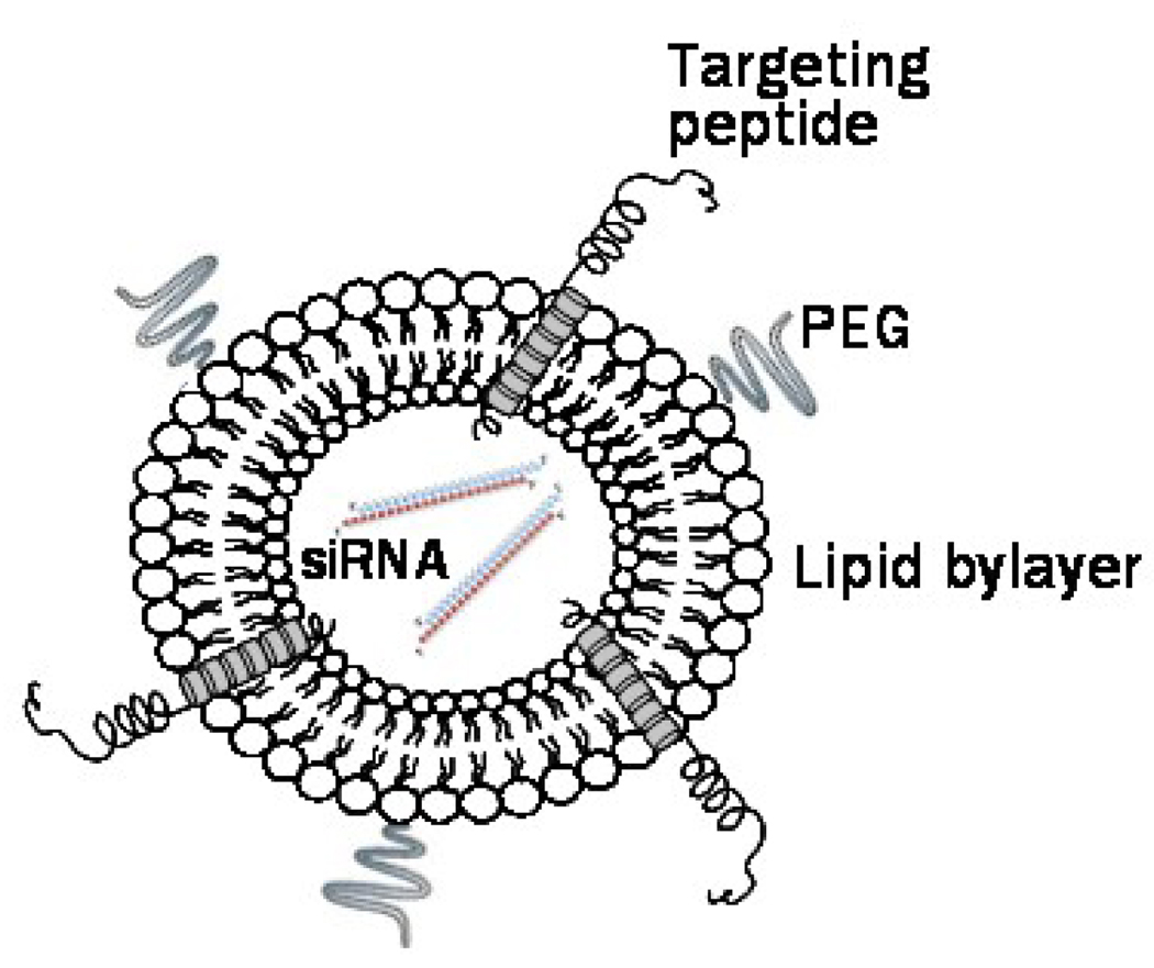
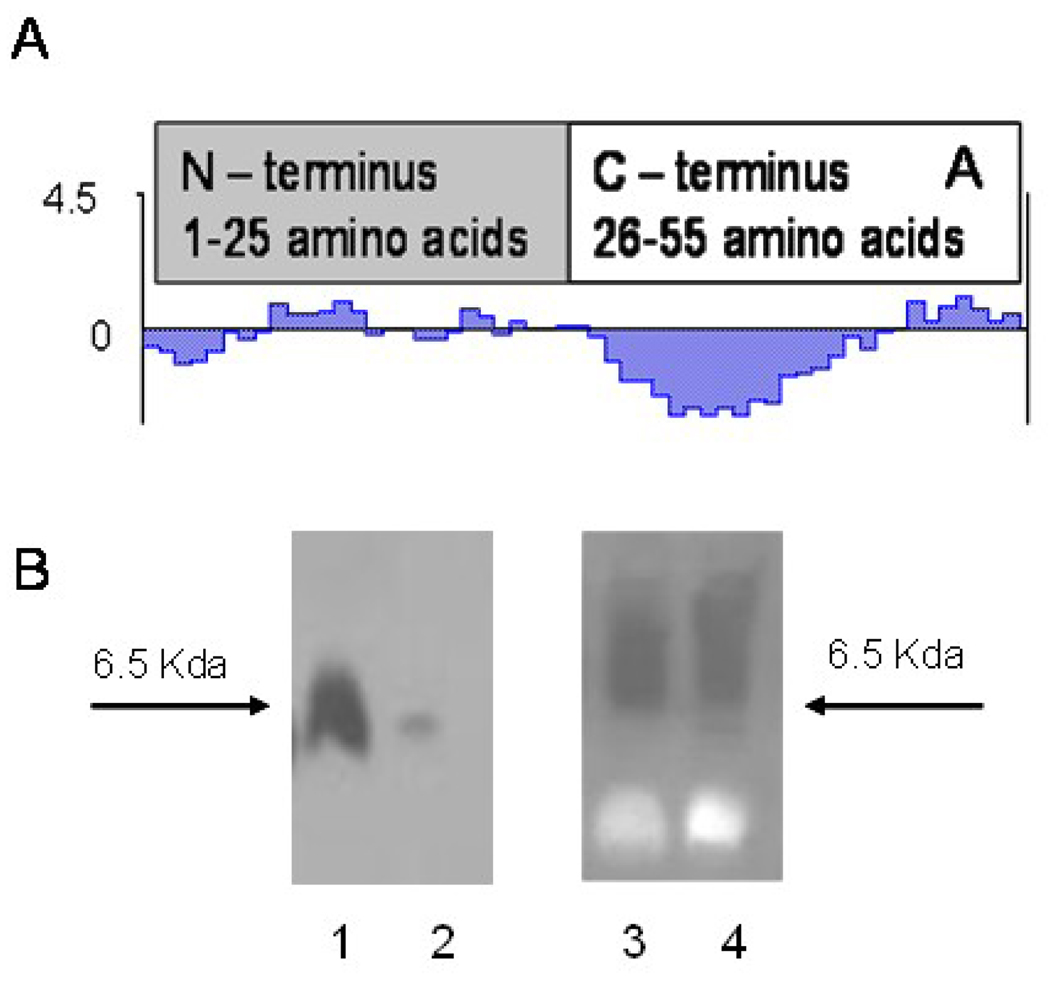
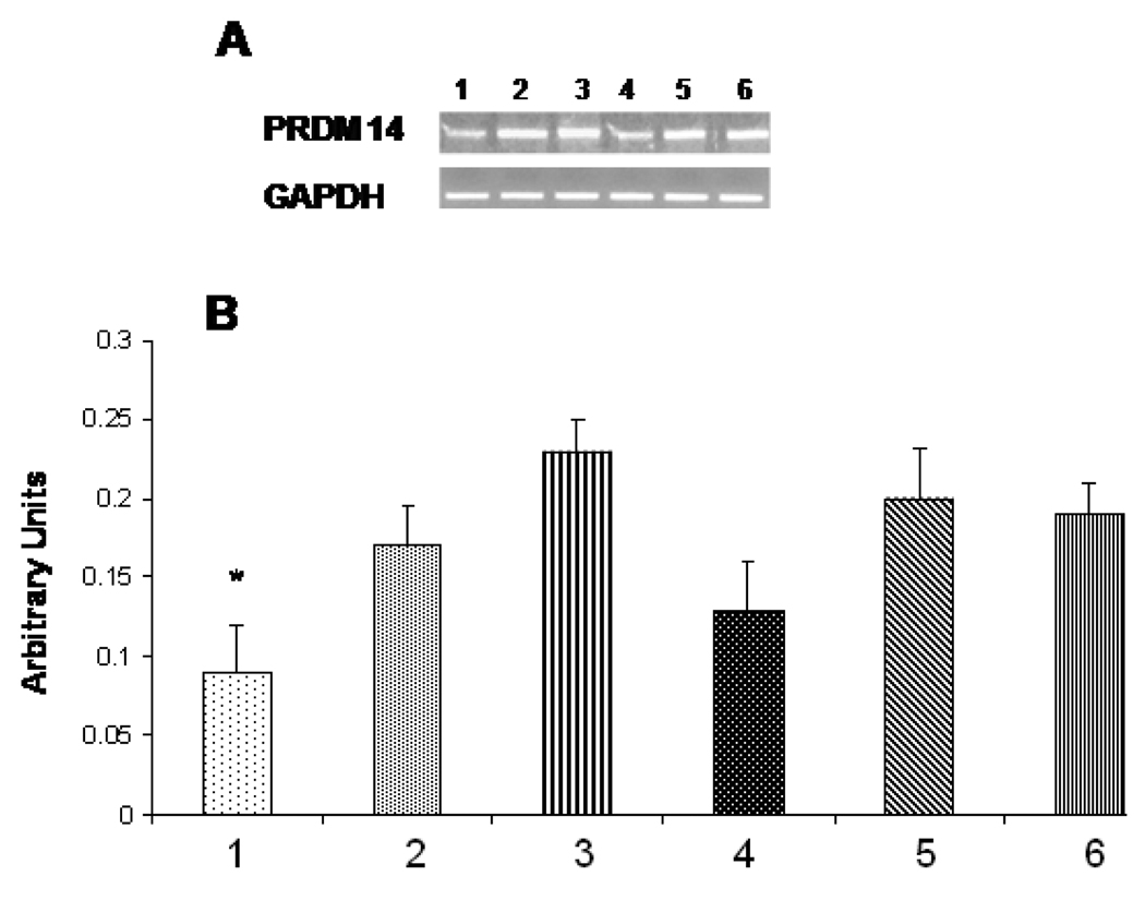
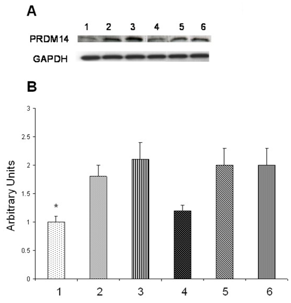
Efficacy of siRNAs as potential anticancer therapeutics can be increased by their targeted delivery into cancer cells via tumor-specific ligands. Phage display offers a unique approach to identify highly specific and selective ligands that can deliver nanocarriers to the site of disease. In this study, we proved a novel approach for intracellular delivery of siRNAs into breast cancer cells through their encapsulation into liposomes targeted to the tumor cells with preselected intact phage proteins. The targeted siRNA liposomes were obtained by a fusion of two parental liposomes containing spontaneously inserted siRNA and fusion phage proteins. The presence of pVIII coat protein fused to a MCF-7 cell-targeting peptide DMPGTVLP in the liposomes was confirmed by Western blotting. The novel phage-targeted siRNA-nanopharmaceuticals demonstrate significant down-regulation of PRDM14 gene expression and PRDM14 protein synthesis in the target MCF-7 cells. This approach offers the potential for development of new anticancer siRNA-based targeted nanomedicines.

From the clinical editor: In this study, the authors report a novel approach for targeted intracellular delivery of siRNAs into breast cancer cells through encapsulation into liposomes targeted to the tumor cells with preselected intact phage proteins.


Conflict of interest statement

There are no disclosures or any conflicts of interest with regard to this publication.

Figures

Figure 1
Figure 1
siRNA-loaded liposome targeted by phage protein fused with a MCF-7 cell-specific peptide DMPGTVLP. The hydrophobic helix of the protein is anchored in the lipid bilayer, whereas the N-terminal fusion peptide DMPGTVLP is displayed on the surface of the liposome. The siRNA molecules are pictured as strands inside the liposomes.
Figure 2
Figure 2
A. Selectivity of phage DMPGTVLP towards breast adenocarcinoma cells MCF-7 in comparison with breast cancer ductal carcinoma cells ZR-75-1, normal breast cells MCF-10A and hepatocellular carcinoma cells HepG2. Phage selectivity was estimated as a percentage phage recovery: output (cell-associated) phage to input phage. The unrelated phage bearing the peptide VPEGAFSS was used as a control. B. Mode of interaction of DMPGTVLP phage with cells MCF-7 under three different conditions (description in the text). The mode of interaction was estimated as a percentage of phage recovery calculated as a ratio of output (cell-associated) phage to input phage. rtp depicts room temperature; sf - serum free medium; s – medium with serum.
Figure 2
Figure 2
A. Selectivity of phage DMPGTVLP towards breast adenocarcinoma cells MCF-7 in comparison with breast cancer ductal carcinoma cells ZR-75-1, normal breast cells MCF-10A and hepatocellular carcinoma cells HepG2. Phage selectivity was estimated as a percentage phage recovery: output (cell-associated) phage to input phage. The unrelated phage bearing the peptide VPEGAFSS was used as a control. B. Mode of interaction of DMPGTVLP phage with cells MCF-7 under three different conditions (description in the text). The mode of interaction was estimated as a percentage of phage recovery calculated as a ratio of output (cell-associated) phage to input phage. rtp depicts room temperature; sf - serum free medium; s – medium with serum.
Figure 3
Figure 3
Mean size (A) and Zeta potential (B) of liposome formulations. Liposomes modified with phage protein (DMPGTVLP-liposomes), liposomes without inserted phage protein (control protein-liposomes), liposomes without inserted siRNA (control siRNA-liposomes) and siRNA liposomes targeted with phage protein (siRNA-DMPGTVLP-liposome) are depicted in the picture.
Figure 4
Figure 4
The presence and topology of phage fusion protein in the protein-modified liposomal preparations determined by Western blot analysis. A. Kyte and Dolittle hydrophillicity (hydropathicity) plot of fusion phage coat protein showing an amphiphilic N-terminus and an intensely hydrophobic segment of the C-terminus. B. Liposomal preparations were treated with proteinase K and then probed with antibodies specific for either N-terminus (Left) or C-terminus (Right) of the phage coat protein. Liposomal association does not provide protection from proteolytic degradation to the N-terminus region of the phage coat protein (Left) but the Cterminus is protected from degradation (Right). Lanes 1, 3 – liposomal preparations untreated with proteinase K, lane 2, 4 – liposomal preparation treated with proteinase K.
Figure 5
Figure 5
Analysis of PRDM14 gene transcription by RT-PCR. MCF-7 cells were treated with PRDM14 gene-specific protein-targeted siRNA–liposomes (40 nM) or protein-targeted scrambled siRNA-liposomes (40 nM) and incubated for 72 h. A. Relative transcription level of the target gene in cells treated with: 1. protein-targeted siRNA–liposomes, 2. protein-targeted scrambled siRNA-liposomes, 3. siRNA-liposomes, 4. siRNA-lipofectamine, 5. Scrambled siRNA-lipofectamine, 6. Control non-treated MCF-7 cells. B. The relative quantification was normalized against GAPDH using KODAK ID image analysis software. All data represent the mean ± S.D. * p<0.05, student-t-test.
Figure 6
Figure 6
Analysis of PRDM14 protein expression by Western blot. MCF-7 cells were treated with PRDM14 gene-specific protein-targeted siRNA–liposomes (40 nM) or protein-targeted scrambled siRNA-liposomes (40 nM) and incubated for 72 h. A. Relative level of protein synthesis in cells treated with: 1. protein-targeted siRNA–liposomes, 2. protein-targeted scrambled siRNA-liposomes, 3. siRNA-liposomes, 4. siRNA-lipofectamine, 5. Scrambled siRNA-lipofectamine, 6. Control non-treated MCF-cells. B. Western blot band intensities quantified using Image J software (NIH). All data represent the mean ± S.D. * p<0.05, student-ttest.

Similar articles

Cited by

References

    1. Iorns E, Lord CJ, Turner N, Ashworth A. Utilizing RNA interference to enhance cancer drug discovery. Nat Rev Drug Discov. 2007;6:556–568. - PubMed
    1. Kim SS, Garg H, Joshi A, Manjunath N. Strategies for targeted nonviral delivery of siRNAs in vivo. Trends in Molecular Medicine. 2009;15:491–500. - PMC - PubMed
    1. Zheng X, Vladau C, Zhang X, Suzuki M, Ichim TE, Zhang ZX, et al. A novel in vivo siRNA delivery system specifically targeting dendritic cells and silencing CD40 genes for immunomodulation. Blood. 2009;113:2646–2654. - PubMed
    1. Pirollo KF, Chang EH. Targeted delivery of small interfering RNA: approaching effective cancer therapies. Cancer Res. 2008;68:1247–1250. - PubMed
    1. Wang W, Tang N, Zhang CL, Liu XJ, Hu H, Zhang ZX, et al. Cell penetrating peptides enhance intracellular translocation and function of siRNA encapsulated in Pegylated liposomes. Yao Xue Xue Bao. 2006;41:142–148. - PubMed

Publication types

MeSH terms