Review
. 2010 Nov;126(5):889-95; quiz 896-7.
doi: 10.1016/j.jaci.2010.09.026.

Innate signals in mucosal immunoglobulin class switching

Affiliations
Review

Innate signals in mucosal immunoglobulin class switching

Irene Puga et al. J Allergy Clin Immunol. 2010 Nov.

Abstract

The intestinal mucosa contains large communities of commensal bacteria that process otherwise indigestible food components, synthesize essential vitamins, stimulate the maturation of the immune system, and form an ecologic niche that prevents the growth of pathogenic species. Conversely, the intestine provides the commensals with a stable habitat rich in energy derived from the ingested food. A delicate homeostatic balance maintains this mutualistic relationship without triggering a destructive inflammatory response. Commensals orchestrate intestinal homeostasis by entertaining an intimate dialogue with epithelial cells and immune cells lodged in the mucosa. Such a dialogue generates finely tuned signaling programs that ensure a state of hyporesponsiveness against noninvasive commensals and a state of active readiness against invasive pathogens. In this dialogue epithelial cells function as "interpreters" that continuously translate microbial messages to "instruct" immune cells as to the antigenic composition of the intestinal lumen. This education process initiates sophisticated defensive strategies that comprise massive production of IgA, a noninflammatory mucosal antibody class that generates immunity while preserving homeostasis.


Figures

FIG 1
FIG 1
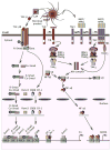
Architecture of intestinal TD and TI IgA responses. Antigen-sampling DCs receive conditioning signals from TLR-activated IECs through TSLP and RA and thereafter differentiate into various DC subsets releasing TGF-β, IL-10, RA, and nitric oxide. These DCs initiate TD IgA responses in Peyer’s patches by inducing TH2, Treg, and Treg-derived TFH cells that activate follicular B cells through CD40L, TGF-β, IL-4, IL-10, and IL-21. IgA-expressing B cells emerging from this TD pathway differentiate into plasma cells in mesenteric lymph nodes and lamina propria. Plasma cells release IgA dimers and IgA oligomers comprising a joining (J) chain that binds to the polymeric immunoglobulin receptor (pIgR) on the basolateral membrane of IECs. This binding triggers endocytosis of polymeric IgA, protease-mediated cleavage of pIgR, and formation of a pIgR-derived secretory component protein that remains associated with the polymeric IgA-J chain complex to form secretory IgA (SIgA). This latter traffics toward the apical membrane of IECs and ultimately gets transcytosed onto the mucosal surface, where it recognizes dietary antigens, commensal bacteria, and pathogens with both high- and low-affinity binding modes. In the lamina propria DCs and IECs can initiate TI IgA responses, including sequential switching from IgA1 to IgA2, by releasing BAFF, APRIL, and RA in response to TLR stimulation by microbial ligands. TSLP from IECs further amplifies IgA production by enhancing DC production of BAFF and APRIL. Together with RA, BAFF and APRIL from DCs and IECs can stimulate plasma cell differentiation and survival in addition to eliciting IgA CSR and production.
FIG 2
FIG 2
Innate IgA-inducing signaling pathways. Mucosal DCs induce IgA CSR and production by releasing BAFF, APRIL, and TGF-β on sensing microbial TLR ligands. Active TGF-κ originates from cleavage of a latency-associated peptide (LAP) by TLR-induced matrix metalloproteases. Engagement of TACI on B cells by BAFF and APRIL triggers association of the adaptor MyD88 to a TACI highly conserved domain that activates NF-κB through a pathway involving IL-1 receptor–associated kinase (IRAK) 1, IRAK-4, TGF-β activated kinase-1 (TAK1), and IκB kinase (IKK)-mediated degradation of the inhibitor of NF-kB (IkB). Additional NF-κB activation involves binding of TNF receptor–associated factor (TRAF) 2 to a TRAF-binding site (TBS) in the cytoplasmic domain of TACI. NF-κB initiates CSR by binding to κB motifs on the AID gene promoter. Engagement of TLRs by microbial ligands enhances IgA CSR and production through a Toll–IL-1 receptor domain (TIR)–dependent pathway that shares MyD88 with the TIR-independent pathway emanating from TACI. Further CSR-inducing signals are provided by TGF-β, which forms a heteromeric TGF receptor (TGF-βR) complex on B cells. TGF-β activates TGF-βR kinases that induce phosphorylation of receptor-regulated SMAD (R-SMAD) proteins. After forming homo-oligomeric and hetero-oligomeric complexes with a common-partner SMAD (Co-SMAD) protein, R-SMAD proteins translocate to the nucleus, where they bind to SMAD-binding elements (SBEs) on the Cαgene promoter. These Smad complexes further associate with constitutive and TGF-βR–induced cofactors, including Runt-related transcription factor 3 (RUNX3), which binds to RUNX-binding elements (RBEs); cyclic adenosine monophosphate response element–binding protein (CREB), which binds to a cyclic adenosine monophosphate response element (CRE); and Ets-like factor-1 (ELF-1), which binds to an Ets-binding site, to enhance CSR from Cμ to Cα.

References

    1. Macpherson AJ, McCoy KD, Johansen FE, Brandtzaeg P. The immune geography of IgA induction and function. Mucosal Immunol. 2008;1:11–22. - PubMed
    1. Cerutti A. The regulation of IgA class switching. Nat Rev Immunol. 2008;8:421–34. - PMC - PubMed
    1. Suzuki K, Maruya M, Kawamoto S, Sitnik K, Kitamura H, Agace WW, et al. The sensing of environmental stimuli by follicular dendritic cells promotes immunoglobulin A generation in the gut. Immunity. 2010;33:71–83. - PubMed
    1. Tsuji M, Suzuki K, Kitamura H, Maruya M, Kinoshita K, Ivanov II, et al. Requirement for lymphoid tissue-inducer cells in isolated follicle formation and T cell-independent immunoglobulin A generation in the gut. Immunity. 2008;29:261–71. - PubMed
    1. Muramatsu M, Nagaoka H, Shinkura R, Begum NA, Honjo T. Discovery of activation-induced cytidine deaminase, the engraver of antibody memory. Adv Immunol. 2007;94:1–36. - PubMed

MeSH terms