Review
. 2011 Jan;24(1-2):11-9.
doi: 10.1093/protein/gzq096. Epub 2010 Nov 3.

Protein folds and protein folding

Affiliations
Review

Protein folds and protein folding

R Dustin Schaeffer et al. Protein Eng Des Sel. 2011 Jan.

Abstract

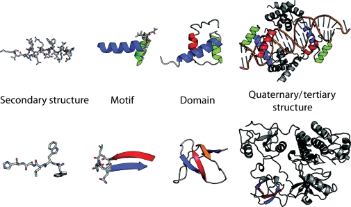
The classification of protein folds is necessarily based on the structural elements that distinguish domains. Classification of protein domains consists of two problems: the partition of structures into domains and the classification of domains into sets of similar structures (or folds). Although similar topologies may arise by convergent evolution, the similarity of their respective folding pathways is unknown. The discovery and the characterization of the majority of protein folds will be followed by a similar enumeration of available protein folding pathways. Consequently, understanding the intricacies of structural domains is necessary to understanding their collective folding pathways. We review the current state of the art in the field of protein domain classification and discuss methods for the systematic and comprehensive study of protein folding across protein fold space via atomistic molecular dynamics simulation. Finally, we discuss our large-scale Dynameomics project, which includes simulations of representatives of all autonomous protein folds.


Figures

Fig. 1
Fig. 1
Structural elements from Pit-1 homeodomain (PDB:1AU7) and Src (PDB:1FMK). Motifs are made up of secondary structure elements but do not necessarily make up a hydrophobic core. Domains are the smallest self-contained unit within a structure. Structures may be made up of multiple domains, sometimes with repeats of the same domain.
Fig. 2
Fig. 2
Structurally similar domains and their structural elements. Some structurally similar proteins contain nearly identical secondary structure elements in similar orientations. Hemoglobin, chain A (PDB:2MHB, chain A) and myoglobin (1A6N) are two such members of the globin fold. Chemotaxis protein Y (PDB:3CHY), Histamine N-methyltransferase (PDB:2AOT) and Catechol O-methyltransferase (1VID) have a conserved structural core surrounded by some non-conserved regions.
Fig. 3
Fig. 3
Domains of guanylate kinase (CATH: 1KGDA01) and translocation ATPase (CATH:1NGDA01), two structurally diverse domains from the CATH v1.73 superfamily 3.40.50.300, the P-loop nucleotide hydrolases. Adapted from Cuff et al. (2009a).
Fig. 4
Fig. 4
Examples of difficult domain partitions. (A) Escherichia coli phosphorin (PDB:1PHO); SCOP and CATH do not partition this structure, DALI partitions it into four separate domains. (B) Periplasmic lysine/arginine/ornithine-binding protein (PDB:2LAO); SCOP does not partition this domain (disfavors discontinuous domains), CATH and DALI partition it into two domains; domain 1 (red/brown), domain 2 (blue). (C) 3-ketoacyl-CoA thiolase (PDB:1PXT), CATH assigns two domains, (D) SCOP assigns two different domains, (E) and the AUTHORS database (Islam et al., 1995) assigns three. Adapted from Veretnik et al. (2004).
Fig. 5
Fig. 5
Major conformational states sampled during thermal unfolding MD simulations. (A) Native (N), transition (TS) and denatured state (D) of CI2. The TS of CI2 is characterized by the packing of the still nascent helix against the partially formed β-sheet. The denatured state of CI2 is particularly denatured, containing little secondary structure. (B) Native (N), transition (TS), intermediate (I) and denatured (D) states of the engrailed homedomain. The TS of EnHD is characterized by essentially native helices condensing into their native topology. The EnHD intermediate contains fully formed helices I and III with a partially denatured helix II. (C) The WW domain does not contain a hydrophobic core. Instead, two small hydrophobic clusters are found on either side of the β-sheet. Residues in cluster 1 (purple) and cluster 2 (cyan), associate in strands 1 (red) and 2 (blue) of FBP28 in the TS. These residues nucleate the folding of the WW domain despite the plasticity of the precise turn residues in the TS.

Similar articles

Cited by

References

    1. Adam G. Protein Sci. 1996;5:1325–1338. - PMC - PubMed
    1. Alexandrov N.N., Go N. Protein Sci. 1994;3:866–875. - PMC - PubMed
    1. Alva V., Koretke K.K., Coles M., Lupas A.N. Curr. Opin. Struct. Biol. 2008;18:358–365. - PubMed
    1. Andreeva A., Howorth D., Chandonia J.M., Brenner S.E., Hubbard T.J., Chothia C., Murzin A.G. Nucleic Acids. Res. 2008;36:D419–D425. - PMC - PubMed
    1. Beck D.A., Jonsson A.L., Schaeffer R.D., Scott K.A., Day R., Toofanny R.D., Alonso D.O., Daggett V. Protein Eng. Des. Sel. 2008;21:353–368. - PubMed

Publication types