Mechanisms of dyspnea
- PMID: 21051395
- PMCID: PMC2972628
- DOI: 10.1378/chest.10-0534
Mechanisms of dyspnea
Abstract
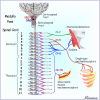
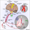
The mechanisms and pathways of the sensation of dyspnea are incompletely understood, but recent studies have provided some clarification. Studies of patients with cord transection or polio, induced spinal anesthesia, or induced respiratory muscle paralysis indicate that activation of the respiratory muscles is not essential for the perception of dyspnea. Similarly, reflex chemostimulation by CO₂ causes dyspnea, even in the presence of respiratory muscle paralysis or cord transection, indicating that reflex chemoreceptor stimulation per se is dyspnogenic. Sensory afferents in the vagus nerves have been considered to be closely associated with dyspnea, but the data were conflicting. However, recent studies have provided evidence of pulmonary vagal C-fiber involvement in the genesis of dyspnea, and recent animal data provide a basis to reconcile differences in responses to various C-fiber stimuli, based on the ganglionic origin of the C fibers. Brain imaging studies have provided information on central pathways subserving dyspnea: Dyspnea is associated with activation of the limbic system, especially the insular area. These findings permit a clearer understanding of the mechanisms of dyspnea: Afferent information from reflex stimulation of the peripheral sensors (chemoreceptors and/or vagal C fibers) is processed centrally in the limbic system and sensorimotor cortex and results in increased neural output to the respiratory muscles. A perturbation in the ventilatory response due to weakness, paralysis, or increased mechanical load generates afferent information from vagal receptors in the lungs (and possibly mechanoreceptors in the respiratory muscles) to the sensorimotor cortex and results in the sensation of dyspnea.
Figures


References
-
- Burki NK. Breathlessness and mouth occlusion pressure in patients with chronic obstruction of the airways. Chest. 1979;76(5):527–531. - PubMed
-
- Burki NK. Dyspnea. Clin Chest Med. 1980;1(1):47–55. - PubMed
-
- Elliott MW, Adams L, Cockcroft A, MacRae KD, Murphy K, Guz A. The language of breathlessness. Use of verbal descriptors by patients with cardiopulmonary disease. Am Rev Respir Dis. 1991;144(4):826–832. - PubMed
-
- Banzett RB, Lansing RW, Reid MB, Adams L, Brown R. ‘Air hunger’ arising from increased PCO2 in mechanically ventilated quadriplegics. Respir Physiol. 1989;76(1):53–67. - PubMed
Publication types
MeSH terms
Grants and funding
LinkOut - more resources
Full Text Sources
Medical

