. 2011 Feb;300(2):G334-44.
doi: 10.1152/ajpgi.00134.2010. Epub 2010 Nov 4.

In vivo analysis of mouse gastrin gene regulation in enhanced GFP-BAC transgenic mice

Affiliations

In vivo analysis of mouse gastrin gene regulation in enhanced GFP-BAC transgenic mice

Shigeo Takaishi et al. Am J Physiol Gastrointest Liver Physiol. 2011 Feb.

Abstract

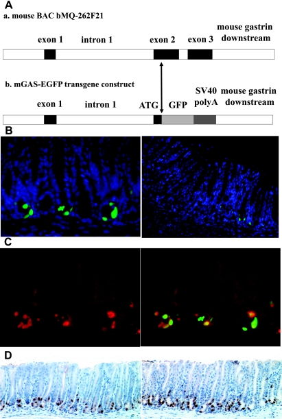
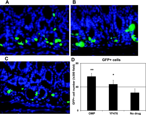
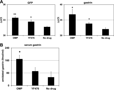
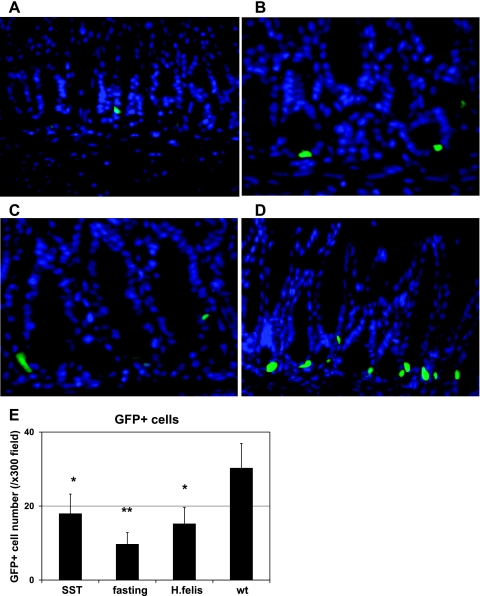
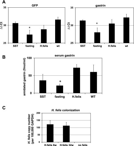
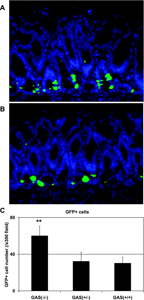
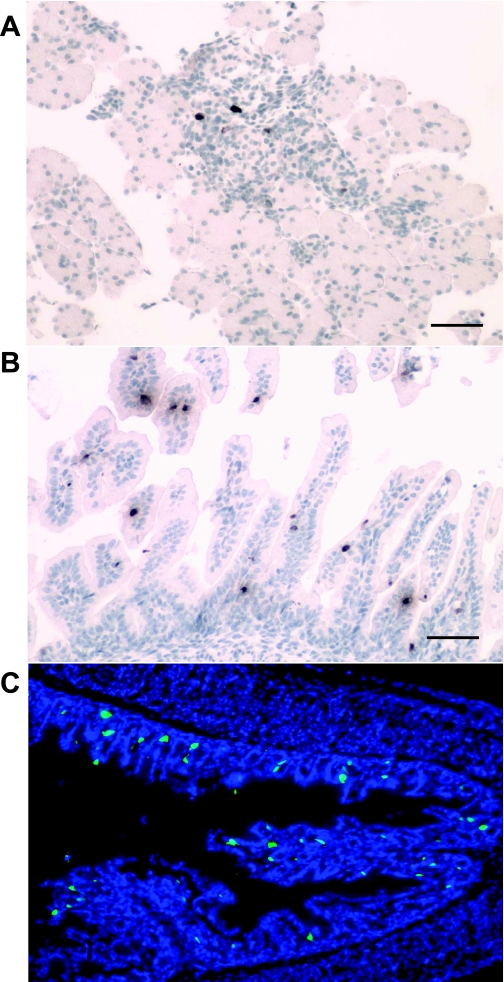
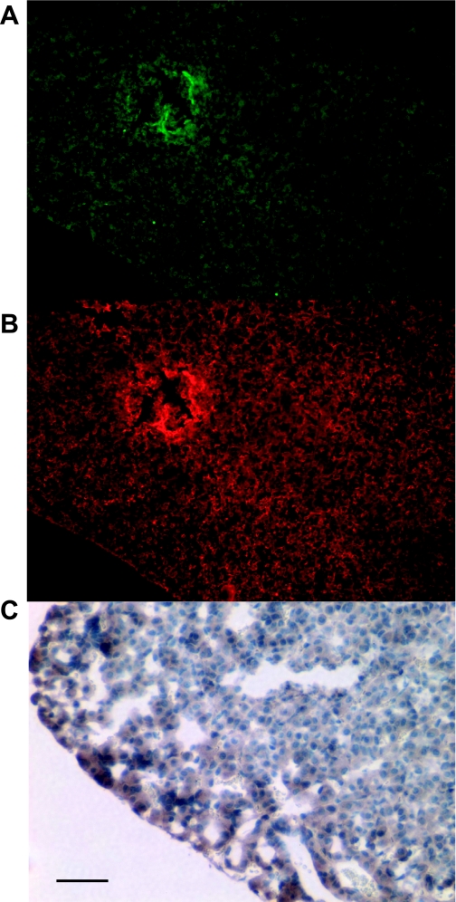
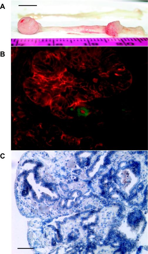
Gastrin is secreted from a subset of neuroendocrine cells residing in the gastric antrum known as G cells, but low levels are also expressed in fetal pancreas and intestine and in many solid malignancies. Although past studies have suggested that antral gastrin is transcriptionally regulated by inflammation, gastric pH, somatostatin, and neoplastic transformation, the transcriptional regulation of gastrin has not previously been demonstrated in vivo. Here, we describe the creation of an enhanced green fluorescent protein reporter (mGAS-EGFP) mouse using a bacterial artificial chromosome that contains the entire mouse gastrin gene. Three founder lines expressed GFP signals in the gastric antrum and the transitional zone to the corpus. In addition, GFP(+) cells could be detected in the fetal pancreatic islets and small intestinal villi, but not in these organs of the adult mice. The administration of acid-suppressive reagents such as proton pump inhibitor omeprazole and gastrin/CCK-2 receptor antagonist YF476 significantly increased GFP signal intensity and GFP(+) cell numbers in the antrum, whereas these parameters were decreased by overnight fasting, octreotide (long-lasting somatostatin ortholog) infusion, and Helicobacter felis infection. GFP(+) cells were also detected in the anterior lobe of the pituitary gland and importantly in the colonic tumor cells induced by administration with azoxymethane and dextran sulfate sodium salt. This transgenic mouse provides a useful tool to study the regulation of mouse gastrin gene in vivo, thus contributing to our understanding of the mechanisms involved in transcriptional control of the gastrin gene.


Figures

Fig. 1.
Fig. 1.
Characterization of the mouse gastrin gene and the transgene construct of bacterial artificial chromosome (BAC) transgenic murine gastrin-promoter enhanced green fluorescent protein (EGFP) reporter (mGAS-EGFP) mice and the expression of green fluorescent protein (GFP) signals in the antrum and the transitional zone of mGAS-EGFP mice. A: genomic segment encompassing the mouse gastrin gene locus is represented as a horizontal line. Exons are represented as filled boxes and introns are unshaded. The transgene construct of mGAS-EGFP mice is created by replacing the open reading frame of the mouse gastrin gene, located in the 2nd and 3rd exons, after the ATG start codon with the EGFP cDNA-SV40polyA-PGK-neomycin cassette by using the BAC recombineering technology, followed by the removal of PGK-neomycin cassette with arabinose-induced Flpe recombinase gene. B: GFP expression in the gastric antrum and corpus of mGAS-EGFP mice. Left: merged image of GFP signals and 4′,6-diamidino-2-phenylindole (DAPI) staining in the gastric antrum (magnification: ×400). Right: merged image of GFP signals and DAPI staining from the gastric corpus to the transitional zone (magnification: ×200). Many GFP-positive cells were detected in the gastric antrum, whereas there were few positive cells in the transitional zone and no positive cells in the corpus. C: immunohistochemical staining with anti-amidated gastrin antibody in the antrum. Anti-amidated gastrin immunostaining with Texas red (left) and merged image of GFP expression with anti-amidated gastrin immunostaining (right) (magnification: ×400). D: bright-field immunohistochemical staining with anti-GFP and anti-amidated gastrin antibodies in the antrum. Anti-GFP antibody immunostaining (left) and anti-amidated gastrin antibody immunostaining (right), both followed by peroxidase-conjugated secondary antibodies and diaminobenzidine staining (magnification: ×200). C and D: images indicated that most of the GFP-positive cells were also positive for gastrin immunostaining, which confirmed that these cells were indeed G cells.
Fig. 2.
Fig. 2.
Increased intensity of GFP signals and GFP-positive cell numbers in the gastric antrum of mGAS-EGFP mice after administration of acid-suppressive reagents. AC: merged images of GFP expression with DAPI staining in the antrum of mGAS-EGFP mice (magnification: ×600). A: mice were injected intraperitoneally with omeprazole (OMP; 5 days per week) for 2 wk. B: mice were injected intraperitoneally with gastrin/CCK2 receptor antagonist YF476 (twice per week) for 2 wk. C: mice were injected intraperitoneally with vehicle only for 2 wk. D: total numbers of GFP-positive cells in the antrum of mGAS-EGFP mice by administration with OMP or YF476 or vehicle only. The number of GFP-positive cells was expressed as the average number of positive cells counted per ×300 magnification high-power field. From each slide, cells were counted from at least 5 fields, and 3 slides (=3 mice) from each group were analyzed. The administration of either OMP or YF476 for 2 wk increased the intensity of GFP signals as well as the number of GFP(+) cells in the gastric antrum of the mGAS-EGFP mice. *P < 0.05, **P < 0.01; n = 3 for each group.
Fig. 3.
Fig. 3.
Quantitative real-time RT-PCR analysis of GFP and gastrin expression in the stomachs of mGAS-EGFP mice and serum gastrin levels after administration of acid-suppressive reagents. A: GFP and gastrin expression were analyzed by quantitative real-time RT-PCR (qRT-PCR) in the stomachs of mGAS-EGFP mice administered with either OMP (5 days per week) or YF476 (twice per week) or vehicle only for 2 wk; n = 3 for each group. Both GFP and gastrin expression in the stomachs of mGAS-EGFP mice treated with either OMP or YF476 were significantly higher than in mice treated with vehicle only. *P < 0.05, **P < 0.01; n = 3 for each group. B: total amidated gastrin levels were measured in serum from mGAS-EGFP mice administered with either OMP (5 days per week) or YF476 (twice per week) or vehicle only for 2 wk; n = 3 for each group. Hypergastrinemia was induced by these acid-suppressive reagents, especially OMP treatment resulted in significantly higher serum gastrin levels compared with vehicle only. *P < 0.05; n = 3 for each group.
Fig. 4.
Fig. 4.
Decreased GFP signal intensity and GFP-positive cell numbers in the gastric antrum of mGAS-EGFP mice following administration of somatostatin, overnight fasting, and Helicobacter felis infection. AD: merged image of GFP expression with DAPI staining in the antrum of mGAS-EGFP mice (magnification: ×600). A: mice were systemically infused with octreotide (long-lasting somatostatin ortholog) for 2 wk by use of osmotic minipumps. B: mice were fasted overnight. C: mice were infected with H. felis for 6 wk. D: mice without treatment (wt) or infection. E: total numbers of GFP-positive cells in the antrum of mGAS-EGFP mice by systemic infusion with the somatostatin ortholog octreotide (SST) for 2 wk, overnight fasting, and H. felis infection for 6 wk. The number of GFP-positive cells were expressed as the average number of positive cells counted per ×300 magnification high-power field. From each slide, cells were counted from at least 5 fields, and 3 slides (=3 mice) from each group were analyzed. The systemic infusion with somatostatin for 2 wk or overnight fasting or H. felis infection for 6 wk decreased the intensity of GFP signals as well as the number of GFP(+) cells in the gastric antrum of the mGAS-EGFP mice. *P < 0.05, **P < 0.01; n = 3 for each group.
Fig. 5.
Fig. 5.
Quantitative real-time RT-PCR analysis of GFP and gastrin expression in the stomachs of mGAS-EGFP mice and serum gastrin levels after systemic infusion of octreotide, overnight fasting, and Helicobacter felis infection. A: GFP and gastrin mRNA expression were analyzed by quantitative real-time RT-PCR in the stomachs of mGAS-EGFP mice after systemic infusion of octreotide, long-lasting somatostatin (SST) homolog, for 2 wk or overnight fasting or Helicobacter felis infection for 6 wk; n = 3 for each group. Both of GFP and gastrin expression in the stomachs of mGAS-EGFP mice after these treatments showed lower expression level, especially overnight fasting resulted in significantly lower expression compared with mice without treatment. *P < 0.05; n = 3 for each group. B: total amidated gastrin levels were measured in serum from mGAS-EGFP mice after systemic infusion of octreotide for 2 wk or overnight fasting or H. felis infection for 6 wk; n = 3 for each group. Hypogastrinemia was induced by octreotide infusion or overnight fasting, especially overnight fasting resulted in significantly lower serum gastrin levels compared with vehicle only, whereas H. felis infection did not change serum gastrin levels. *P < 0.05; n = 3 for each group. C: H. felis colonization in the stomachs of H. felis-infected mGAS-EGFP transgenic mice at 6 and 32 wk (6w and 32w) postinfection as measured by quantitative real-time PCR of genomic DNAs; n = 3 for each group. DNA copy numbers of H. felis normalized to GAPDH (per 100,000 copies) in the stomachs were approximately the same among the 2 time points. Of note, no H. felis DNAs were detected in the stomachs of mGAS-EGFP mice without infection.
Fig. 6.
Fig. 6.
Increase in the intensity of GFP signals and GFP-positive cell numbers in the gastric antrum of gastrin-deficient (GAS-KO)/mGAS-EGFP double-mutant mice. A: merged image of GFP expression with DAPI staining in the antrum of homozygous GAS-KO/mGAS-EGFP double-mutant mice. B: merged image of GFP expression with DAPI staining in the antrum of heterozygous GAS-KO/mGAS-EGFP double-mutant mice (magnification for A and B: ×400). C: total numbers of GFP-positive cells in the antrum of homozygous (GAS−/−) and heterozygous (GAS+/−) GAS-KO/mGAS-EGFP double-mutant mice and mGAS-EGFP single-transgenic mice (GAS+/+). The number of GFP-positive cells were expressed as the average number of positive cells counted per ×300 magnification high-power field. From each slide, cells were counted from at least 5 fields, and 3 slides (=3 mice) from each group were analyzed. GFP(+) cell numbers in the gastric antrum were significantly increased in homozygous GAS-KO/mGAS-EGFP double-mutant mice compared with heterozygous GAS-KO/mGAS-EGFP double-mutant or mGAS-EGFP single-transgenic mice. **P < 0.01; n = 3 for each group.
Fig. 7.
Fig. 7.
GFP expression in the fetal and neonatal organs of mGAS-EGFP mice. A and B: immunohistochemical staining with anti-GFP antibody in the fetal pancreas (A) and small intestinal villi (B) of mGAS-EGFP mice at 18.5 days postconception (dpc). C: merged image of GFP expression with DAPI staining in the gastric antrum of neonatal mGAS-EGFP mice at 2 days after birth (magnification for AC: ×200; scale bar for A and B: 50 μm). GFP(+) cells were detected in the pancreatic islets and the small intestinal villi of the fetus of mGAS-EGFP mice at 18.5 dpc, whereas GFP(+) cells appeared in the gastric antrum of neonatal mGAS-EGFP mice at 2 days after birth. Of note, GFP(+) cells in the neonatal antrum were located at not only the base of the antral glands but also higher up in the middle to the top third of the glands.
Fig. 8.
Fig. 8.
GFP expression in the anterior lobe of the pituitary gland of mGAS-EGFP mice. A: GFP expression in the anterior lobe of the pituitary gland of mGAS-EGFP mice. B: immunofluorescence staining with anti-amidated gastrin antibody followed by Texas red-conjugated anti-goat secondary antibody in the same slides as A (magnification for AB: ×100). C: immunohistochemistry with anti-GFP antibody in the anterior lobe of the pituitary gland of mGAS-EGFP mice (scale bar: 50 μm, magnification: ×300). Weak GFP signals were observed in the anterior lobe of the pituitary gland of mGAS-EGFP mice, and immunofluorescence staining with anti-amidated gastrin antibody confirmed that these GFP-positive cells in the anterior lobe of the pituitary gland were also positive for amidated gastrin.
Fig. 9.
Fig. 9.
GFP expression in the cancer cells of the colon tumors induced by administration with azoxymethane (AOM) and dextran sodium sulfate (DSS) in hyperprogastrinemic hGAS/mGAS-EGFP double-transgenic mice. A: macroimage of the colorectal tumors of hGAS/mGAS-EGFP double-transgenic mice after 4 mo of administration with AOM and DSS (scale bar: 5 mm). B: merged image of GFP expression with fluorescence immunostaining with anti-EpCAM antibody followed by Texas red-conjugated anti-rat secondary antibody in the colorectal tumors of hGAS/mGAS-EGFP mice after 4 mo of administration with AOM and DSS (magnification: ×600). C: immunohistochemistry with anti-GFP antibody in the colon tumors of hGAS/mGAS-EGFP mice after 4 mo of administration with AOM and DSS (scale bar: 50 μm, magnification: ×200). Most of the GFP(+) cells were detected in the neoplastic colonic glands, and they were EpCAM positive, which indicated they were epithelial cancer cells.

Similar articles

Cited by

References

    1. Aly A, Shulkes A, Baldwin GS. Gastrins, cholecystokinins and gastrointestinal cancer. Biochim Biophys Acta 1704: 1– 10, 2004 - PubMed
    1. Brand SJ, Fuller PJ. Differential gastrin gene expression in rat gastrointestinal tract and pancreas during neonatal development. J Biol Chem 263: 5341– 5347, 1988 - PubMed
    1. Brand SJ, Stone D. Reciprocal regulation of antral gastrin and somatostatin gene expression by omeprazole-induced achlorhydria. J Clin Invest 82: 1059– 1066, 1988 - PMC - PubMed
    1. Caplin M, Khan K, Savage K, Rode J, Varro A, Michaeli D, Grimes S, Brett B, Pounder R, Dhillon A. Expression and processing of gastrin in hepatocellular carcinoma, fibrolamellar carcinoma and cholangiocarcinoma. J Hepatol 30: 519– 526, 1999 - PubMed
    1. Caplin M, Savage K, Khan K, Brett B, Rode J, Varro A, Dhillon A. Expression and processing of gastrin in pancreatic adenocarcinoma. Br J Surg 87: 1035– 1040, 2000 - PubMed

Publication types

MeSH terms

LinkOut - more resources