. 2011 Jan 14;286(2):1508-16.
doi: 10.1074/jbc.M110.162966. Epub 2010 Nov 4.

ZFP260 is an inducer of cardiac hypertrophy and a nuclear mediator of endothelin-1 signaling

Affiliations

ZFP260 is an inducer of cardiac hypertrophy and a nuclear mediator of endothelin-1 signaling

Hiba Komati et al. J Biol Chem. .

Abstract

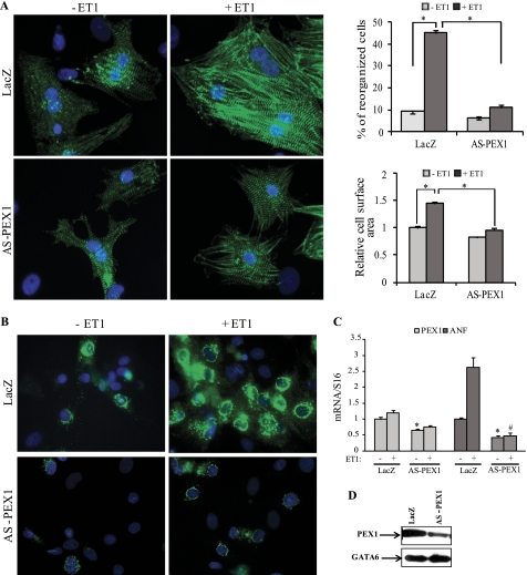
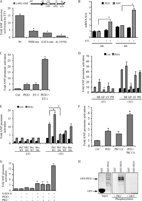
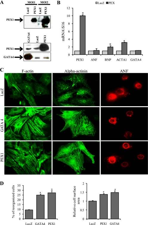
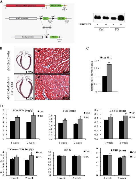
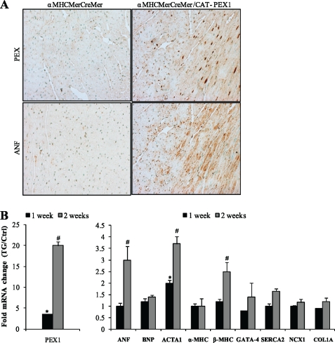
Pressure and volume overload induce hypertrophic growth of postnatal cardiomyocytes and genetic reprogramming characterized by reactivation of a subset of fetal genes. Despite intense efforts, the nuclear effectors of cardiomyocyte hypertrophy remain incompletely defined. Endothelin-1 (ET-1) plays an important role in cardiomyocyte growth and is involved in mediating the neurohormonal effects of mechanical stress. Here, we show that the phenylephrine-induced complex-1 (PEX1), also known as zinc finger transcription factor ZFP260, is essential for cardiomyocyte response to ET-1 as evidenced in cardiomyocytes with PEX1 knockdown. We found that ET-1 enhances PEX1 transcriptional activity via a PKC-dependent pathway which phosphorylates the protein and further potentiates its synergy with GATA4. Consistent with a role for PEX1 in cardiomyocyte hypertrophy, overexpression of PEX1 is sufficient to induce cardiomyocyte hypertrophy in vitro and in vivo. Importantly, transgenic mice with inducible PEX1 expression in the adult heart develop cardiac hypertrophy with preserved heart function. Together, the results identify a novel nuclear effector of ET-1 signaling and suggest that PEX1 may be a regulator of the early stages of cardiac hypertrophy.


Figures

FIGURE 1.
FIGURE 1.
PEX1 is required for ET-1 signaling. A, left, Hoechst (blue) and sarcomeric α-actinin (green) immunofluorescence-labeled slides are shown of the control adeno-LacZ and adeno-HA-AS-PEX1-infected primary cardiomyocytes treated or not with 100 nm ET-1 for 24 h. Costaining was performed using Hoechst to detect cell nuclei and sarcomeric α-actinin to visualize the myofibrils of cardiomyoctes. Note that the loss of PEX1 blocked the myofibrillar reorganization seen in adeno-LacZ-infected cells treated with ET-1. A, right, reorganized cells were counted, and relative cell surface area was measured across 10 fields (×40) in three separate experiments. *, p < 0.05. B, ANF immunofluorescence is shown in cardiomyocytes infected for 3 days with either adeno-LacZ or adeno-HA-AS-PEX1 and treated with vehicle or ET-1. Note how PEX1 down-regulation inhibited ET-1-induced cellular accumulation of ANF. C, ANF mRNA levels in LacZ- or AS-PEX1-transfected cardiomyocytes treated or not with ET-1. *, p < 0.05 versus LacZ; #, p < 0.05 versus LacZ + ET-1. D, PEX1 protein levels in cardiomyocytes infected with the adeno-AS-PEX1 or adeno-LacZ as detected by Western blotting are shown. GATA6 protein was used as an internal control.
FIGURE 2.
FIGURE 2.
Post-translational regulation of PEX1 by ET-1. A, ANF promoter elements required for ET-1 response in cardiomyocytes. Cells were transfected with wild-type (Wt) and mutated −695ANF or −135ANF promoter luciferase reporter constructs and stimulated for 48 h with 100 nm ET-1. PERE mut corresponds to mutation of the proximal PERE site (position −70 bp); GATA mut corresponds to mutation of the GATA site (position −120 bp). The data are mean of ± S.E. (error bars; n = 4). *, p < 0.05 versus wild type. B, effect of ET-1 on PEX1 mRNA levels. ANF and PEX1 transcript levels were quantified in primary cultured ventricular cardiomyocytes treated or not with 100 nm ET-1 for 24 or 48 h. Data are mean ± S.E. (n = 4). *, p < 0.05 C, ET-1 potentiates PEX1 activation. Cardiomyocytes were cotransfected with the proximal ANF promoter luciferase reporter and 500 ng of PEX1. The cells were then treated with 100 nm ET-1 for 48 h. Data are mean ± S.E. (n = 4). *, p < 0.05 versus Ctrl. D, involvement of PKC but not p38-MAPK or ERK1/2 in ET-1-dependent PEX1 activation of ANF transcription. Cardiomyocytes were cotransfected with the proximal ANF promoter luciferase reporter and 500 ng of PEX1. The cells were then treated with 100 nm ET-1 for 24 h in the presence of inhibitors: p38 MAPK (SB 203580; 10 μm [SB]), PKC (GF 109203X; 5 μm [GF]), PI3K (LY 294002; 25 μm [LY]) ERK1/2 (PD98059; 10 μm [PD]). The data are from one representative experiment carried out in triplicate. *, p < 0.05 versus PEX1 (−ET1); #, p < 0.05 versus PEX1 (+ET1). E and F, cotransfection in cardiomyocytes of the ANF-Luc reporter with the indicated expression vectors treated or not with 100 nm ET-1(PKC CA, PKCβ catalytic domain). Note how cotransfection with PKC-dominant negative (DN) but not Rho-DN abrogates the ET-1 effect on PEX1 activation of the promoter. *, p < 0.05. G, PKCβ synergizes with PEX1 and GATA4 on the ANF promoter. NIH3T3 cells were cotransfected with the ANF-Luc construct 100 ng of PEX1, 10 ng of GATA4, and 200 ng of PKCβ CA. *, p < 0.05 versus Ctrl. H, in vitro PKC phosphorylation of GST-PEX1 fusion protein. In similar kinase assays (such as AKT kinase), PEX1 was not phosphorylated, demonstrating PKC specificity. Coomassie staining was used to show protein loading.
FIGURE 3.
FIGURE 3.
Overexpression of PEX1 is sufficient to induce myocyte hypertrophy. A, upper, representative Western blot confirming overexpression of PEX1 in adeno-HA-PEX1-infected cardiomyocytes. A, lower, Western blot showing no change in PEX1 levels in cardiomyocytes overexpressing GATA4 and no change in GATA4 levels in myocytes overexpressing PEX1. B, histograms showing mRNA fold change of PEX1, ANF, BNP, ACTA1, and GATA4 in adeno-LacZ- or adeno-HA-PEX1-infected cardiomyocytes. The data are expressed as fold change relative to LacZ with S16 as the normalizer gene. Overexpression of PEX1 caused an up-regulation of BNP and ACTA1 mRNA. *, p < 0.05 versus LacZ. C, F-actin detected with the phalloidin-Alexa Fluor 488 (green in left panel), Sarcomeric α-actinin (green in middle panel) and ANF (red in right panel) immunofluoresence-labeled adeno-LacZ-, adeno-GATA4-, or adeno-HA-PEX1-infected cardiomyocytes. Note how induction of either GATA4 or PEX1 induces myofibrillar reorganization (as shown in both the phalloidin and the α-actinin staining) but not ANF accumulation. D, reorganized cells were counted and relative cell surface area measured across 10 fields (×40) in three separate experiments. *, p < 0.05 versus LacZ.
FIGURE 4.
FIGURE 4.
Conditional overexpression of PEX1 in adult mice hearts leads to cardiac hypertrophy. A, strategy used for conditional expression of PEX1 using the Cre/LoxP system. α-MHCMerCreMer mice were crossed with CAT-PEX1 transgenic mice to obtain a conditional transgenic line that overexpresses PEX1 strictly in cardiomyocytes once Tamoxifen is administered. Right, transgenic mice receiving tamoxifen showing increased PEX1 protein levels, whereas those injected with peanut oil and controls receiving tamoxifen showing no change. B, image magnified ×1.25 of trichrome-stained heart sections from 150-day-old α-MHCMerCreMer or α-MHCMerCreMer/CAT-PEX1 mice treated with Tamoxifen. Notice the hypertrophied heart when PEX1 expression is induced. Magnification ×63 shows increased myocyte size. C, relative cell surface area was measured across 10 fields (×40) in transgenic (TG) and control (Ctrl) mice (2 of each). *, p < 0.05 versus control. D, echocardiographic analysis of mice hearts 1 week and 2 weeks after Tamoxifen administration. Heart weight-to-body weight ratios (HW/BW) are also shown. IVS, interventricular septum thickness; LVPW, left ventricular posterior wall thickness; LVID, left ventricular internal dimension; LV mass, left ventricular mass; EF, ejection fraction. Notice how ejection fraction was not changed despite the increase in left ventricular mass/body weight ratio suggestive of preserved heart function. The data are means ± S.E. (error bars; n = 4–5 for each group). *, p < 0.05 versus control (Ctrl; 1 week); #, p < 0.05 versus control (2 weeks).
FIGURE 5.
FIGURE 5.
Genetic changes in PEX1-expressing hearts. A, histologic sections stained with either the PEX1 antibody or ANF antibody and counterstained with methyl green. Note the increased nuclear distribution of PEX1 and the concomitant up-regulation of ANF in the Tamoxifen-treated transgenics versus Tamoxifen-treated controls. B, -fold change in transcript levels relative to controls for PEX1, ANF, BNP, ACTA1, α-MHC, β-MHC, GATA4, SERCA2, NCX1, and COL1A1 in hearts of transgenic mice after 1 and 2 weeks of Tamoxifen treatment (ratio of transgenic (TG) to controls (Ctrl)). The controls of 1 and 2 weeks of Tamoxifen treatment were set at 1.The data are means ± S.E. (error bars; n = 4–5 for each group). S16 was used as the normalizer gene. *, p < 0.05 versus control (1 week); #, p < 0.05 versus control (2 weeks).

Similar articles

Cited by

References

    1. MacLellan W. R., Schneider M. D. (2000) Annu. Rev. Physiol. 62, 289–319 - PubMed
    1. Olson E. N., Schneider M. D. (2003) Genes Dev. 17, 1937–1956 - PubMed
    1. Frey N., Olson E. N. (2003) Annu. Rev. Physiol. 65, 45–79 - PubMed
    1. Ito H., Hirata Y., Adachi S., Tanaka M., Tsujino M., Koike A., Nogami A., Murumo F., Hiroe M. (1993) J. Clin. Invest. 92, 398–403 - PMC - PubMed
    1. Iwanaga Y., Kihara Y., Hasegawa K., Inagaki K., Yoneda T., Kaburagi S., Araki M., Sasayama S. (1998) Circulation 98, 2065–2073 - PubMed

Publication types

LinkOut - more resources