. 2011 Jan;155(1):354-69.
doi: 10.1104/pp.110.166546. Epub 2010 Nov 4.

Plastids contain a second sec translocase system with essential functions

Affiliations

Plastids contain a second sec translocase system with essential functions

Courtney A Skalitzky et al. Plant Physiol. 2011 Jan.

Abstract

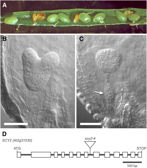
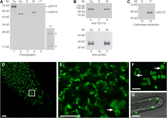
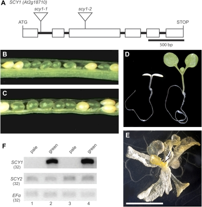
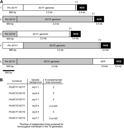
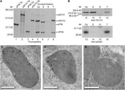
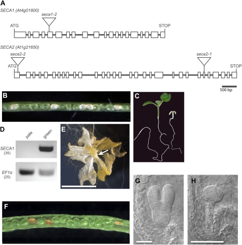
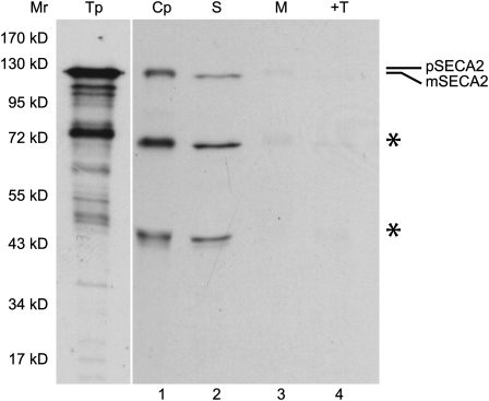
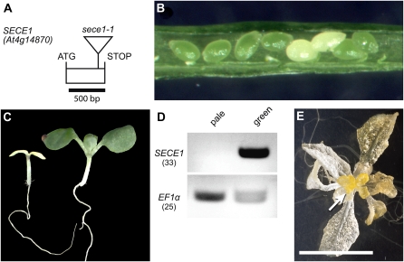
Proteins that are synthesized on cytoplasmic ribosomes but function within plastids must be imported and then targeted to one of six plastid locations. Although multiple systems that target proteins to the thylakoid membranes or thylakoid lumen have been identified, a system that can direct the integration of inner envelope membrane proteins from the stroma has not been previously described. Genetics and localization studies were used to show that plastids contain two different Sec systems with distinct functions. Loss-of-function mutations in components of the previously described thylakoid-localized Sec system, designated as SCY1 (At2g18710), SECA1 (At4g01800), and SECE1 (At4g14870) in Arabidopsis (Arabidopsis thaliana), result in albino seedlings and sucrose-dependent heterotrophic growth. Loss-of-function mutations in components of the second Sec system, designated as SCY2 (At2g31530) and SECA2 (At1g21650) in Arabidopsis, result in arrest at the globular stage and embryo lethality. Promoter-swap experiments provided evidence that SCY1 and SCY2 are functionally nonredundant and perform different roles in the cell. Finally, chloroplast import and fractionation assays and immunogold localization of SCY2-green fluorescent protein fusion proteins in root tissues indicated that SCY2 is part of an envelope-localized Sec system. Our data suggest that SCY2 and SECA2 function in Sec-mediated integration and translocation processes at the inner envelope membrane.


Figures

Figure 1.
Figure 1.
scy2 mutant allele and mutant phenotypes. A, Silique from a SCY2/scy2-4 heterozygous parent showing aborted seeds. B and C, Seeds in younger, developing siliques contain either wild-type-appearing embryos (B) or embryos arrested at the globular stage (C). Extra cell divisions in the suspensor are denoted by the arrow. D, Gene diagram showing introns and exons (boxes) in the open reading frame of SCY2 (At2g31530) and the location of the T-DNA insertion in scy2-4.
Figure 2.
Figure 2.
Arabidopsis SCY2 is a membrane-integrated plastid protein. A to C, In vitro import assays. A, Radiolabeled precursor to SCY2 (pSCY2) was incubated with isolated pea chloroplasts in an in vitro import reaction for 30 min. Chloroplasts were treated with 100 μg mL−1 thermolysin, repurified, washed, lysed, and fractionated into stroma and membranes. Translation product (Tp) equivalent to 5% of the assay, chloroplasts (Cp), stroma (S), membranes (M), and thermolysin-treated membranes (+T) were analyzed using SDS-PAGE and fluorography. A longer exposure (4×) of thermolysin-treated membranes is shown at right to visualize mature SCY2 degradation products, indicated by asterisks. B, Chloroplasts, stroma, and membranes were also analyzed by SDS-PAGE and immunodetection with antibodies to TIC110 (applied at 1:5,000) or cpTatC (applied at 1:20,000). C, Membranes from the SCY2 import reaction were subjected to alkaline carbonate extraction. The supernatant (S) and membrane pellet (P) were analyzed using SDS-PAGE and fluorography. The positions of molecular mass markers (Mr) are indicated at the left of each gel or blot image. D to G, Localization of the SCY2-GFP fusion protein in root tips (D–F) and root hairs (G) by confocal laser scanning microscopy. The boxed area in D indicates the amyloplasts in the columella of the root tip. E and F are higher magnification views showing plastids of different morphology. Amyloplasts in the columella cells are indicated by the white arrow in E, and stromules are indicated by the white arrows in F. Bars = 10 μm.
Figure 3.
Figure 3.
scy1 mutant alleles and mutant phenotypes. A, Gene diagram showing introns and exons (boxes) in the open reading frame of SCY1 (At2g18710) and the location of T-DNA insertions in scy1-1 and scy1-2. B and C, Opened siliques showing white seeds in scy1-1 (B) and scy1-2 (C). D, Pale homozygous scy1-1 mutant seedling and green sibling. E, scy1-1 plant grown for 5 weeks on 1% Suc. The arrow indicates floral buds. Bar = 1 cm. F, RT-PCR analyses. cDNA prepared from RNA isolated from either pale seedlings or green siblings was amplified with primers flanking the T-DNA insertion sites. Lanes 1 and 2, scy1-1; lanes 3 and 4, scy1-2. The number of PCR cycles is indicated by parentheses. Full-length SCY1 transcripts do not accumulate in the pale seedlings.
Figure 4.
Figure 4.
Complementation of scy mutants. A, Constructs used for SCY mutant complementation, promoter-swap experiments, and localization experiments. B, Table indicating the genetic backgrounds and number of independent lines where complementation was confirmed for each construct. For backgrounds where complementation was not observed, the number of lines that were screened is shown.
Figure 5.
Figure 5.
SCY2 localizes to the envelope membranes. A, Import assays followed by membrane fractionation. Radiolabeled precursors to P36 (pP36) and SCY2 (pSCY2) were each incubated with isolated pea chloroplasts in in vitro import reactions for 15 min. Both precursors were also coimported into isolated chloroplasts for 15 min. Chloroplasts from all three import reactions were repurified and washed. Chloroplasts that underwent coimport were lysed and fractionated (see “Materials and Methods”). Translation products (Tp) equivalent to 5% of each assay, chloroplasts (Cp), envelope membranes (E), stroma (S), and thylakoid membranes (T) were analyzed by SDS-PAGE and fluorography. B, Immunoblots of chloroplasts and fractions from pSCY2/pP36 coimport probed with antibodies to TIC110 (applied at 1:5,000) or cpTatC (applied at 1:20,000). C to E, Immunogold localization of GFP. Thin sections of plastids in root cells expressing SCY2-GFP (C and D) or wild-type root cells (E) were incubated with GFP antibodies, applied at 1:10 (C) or 1:30 (D and E), and goat anti-rabbit secondary antibodies conjugated to 10-nm gold (C) or 15-nm gold (D and E). The gold label is found preferentially at the periphery of the plastids. Bars = 500 nm.
Figure 6.
Figure 6.
seca mutant alleles and mutant phenotypes. A, Gene diagrams of SECA1 (At4g01800) and SECA2 (At1g21650) showing introns and exons (boxes) in the open reading frames and the location of T-DNA insertions in the mutants. B, Opened silique of a SECA1/seca1-2 heterozygous plant showing white seeds. C, Pale seca1-2 homozygous mutant seedling and green sibling. D, RT-PCR analysis. cDNA prepared from RNA isolated from either pale seedlings or green siblings was amplified with primers flanking the seca1-2 T-DNA insertion site or primers specific for EF1α (loading control). The number of PCR cycles is indicated by parentheses. Full-length SECA1 transcripts do not accumulate in the pale seedlings. E, A seca1-2 plant grown for 5 weeks on 1% Suc. The arrow indicates floral buds. Bar = 1 cm. F, Opened silique of a SECA2-1/seca2-1 heterozygous plant showing aborted seeds. G and H, Seeds in younger, developing siliques contain either wild-type-appearing embryos (G) or embryos arrested at the globular stage (H). Bars = 50 μm.
Figure 7.
Figure 7.
Arabidopsis SECA2 is a plastid-localized protein. Radiolabeled precursor to SECA2 (pSECA2), obtained by a coupled transcription and translation reaction, was incubated with isolated chloroplasts in an in vitro import reaction for 45 min. Chloroplasts were treated with 100 μg mL−1 thermolysin, repurified, washed, lysed, and fractionated into stroma and membranes. Translation product (Tp) equivalent to 0.8% of the assay, chloroplasts (Cp), stroma (S), membranes (M), and thermolysin-treated membranes (+T) were analyzed using SDS-PAGE and fluorography. Smaller translation products, likely to be carboxyl-truncated pSECA2 resulting from premature termination, also appeared to be imported and processed, as indicated by asterisks. Translation products that were exposed for one-fourth the length of time of chloroplasts and chloroplast fractions are shown. The positions of molecular mass markers (Mr) are shown at left.
Figure 8.
Figure 8.
sece1 mutant allele and mutant phenotypes. A, Gene diagram of SECE1 showing the single exon in the open reading frame and the location of the T-DNA insertion in the sece1-1 mutant. B, Opened silique of a SECE1/sece1-1 heterozygous plant showing white seeds. C, Pale sece1-1 homozygous mutant seedling and green sibling. D, RT-PCR analysis. cDNA prepared from RNA isolated from either pale seedlings or green siblings was amplified with primers flanking the sece1-1 T-DNA insertion site or primers specific for EF1α. The number of PCR cycles is indicated by parentheses. Full-length SECE1 transcripts do not accumulate in the pale seedlings. E, sece1-1 plant grown for 5 weeks on 1% Suc. The arrow indicates floral buds. Bar = 1 cm.

Similar articles

Cited by

References

    1. Adamczyk BJ, Lehti-Shiu MD, Fernandez DE. (2007) The MADS domain factors AGL15 and AGL18 act redundantly as repressors of the floral transition in Arabidopsis. Plant J 50: 1007–1019 - PubMed
    1. Alonso JM, Stepanova AN, Leisse TJ, Kim CJ, Chen H, Shinn P, Stevenson DK, Zimmerman J, Barajas P, Cheuk R, et al. (2003) Genome-wide insertional mutagenesis of Arabidopsis thaliana. Science 301: 653–657 - PubMed
    1. Andersson H, von Heijne G. (1993) Sec dependent and Sec independent assembly of E. coli inner membrane proteins: the topological rules depend on chain length. EMBO J 12: 683–691 - PMC - PubMed
    1. Benz M, Bals T, Gügel IL, Piotrowski M, Kuhn A, Schünemann D, Soll J, Ankele E. (2009) Alb4 of Arabidopsis promotes assembly and stabilization of a non chlorophyll-binding photosynthetic complex, the CF1CF0-ATP synthase. Mol Plant 2: 1410–1424 - PubMed
    1. Berghöfer J, Karnauchov I, Herrmann RG, Klösgen RB. (1995) Isolation and characterization of a cDNA encoding the SecA protein from spinach chloroplasts: evidence for azide resistance of Sec-dependent protein translocation across thylakoid membranes in spinach. J Biol Chem 270: 18341–18346 - PubMed

Publication types