. 2011 Mar 14;52(3):1368-77.
doi: 10.1167/iovs.10-6125.

Factors impacting corneal epithelial barrier function against Pseudomonas aeruginosa traversal

Affiliations

Factors impacting corneal epithelial barrier function against Pseudomonas aeruginosa traversal

Irania Alarcon et al. Invest Ophthalmol Vis Sci. .

Abstract

Purpose: Mechanisms determining epithelial resistance versus susceptibility to microbial traversal in vivo remain poorly understood. Here, a novel murine model was used to explore factors influencing the corneal epithelial barrier to Pseudomonas aeruginosa penetration.

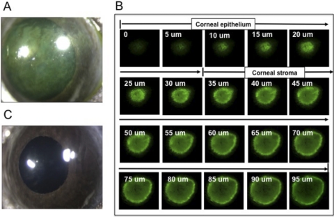
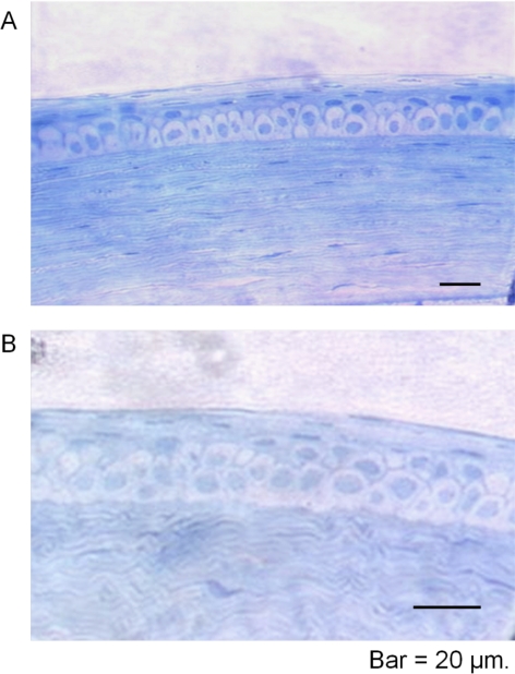
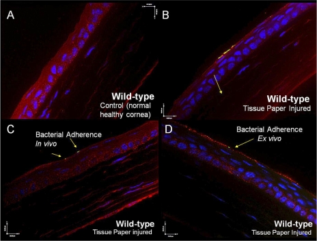
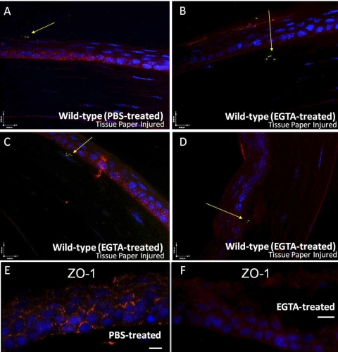
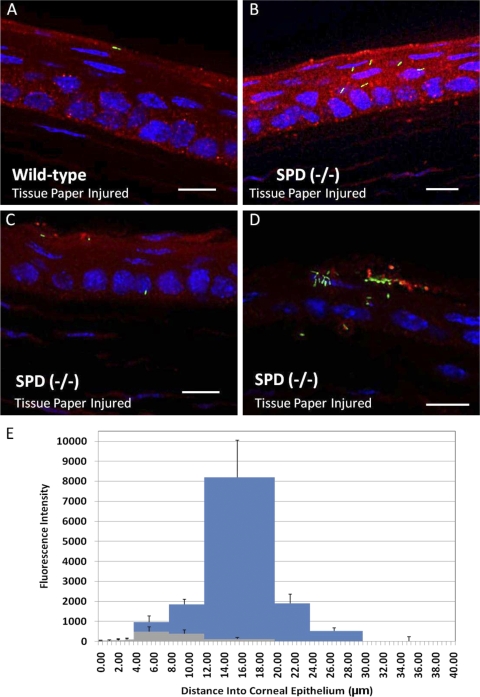
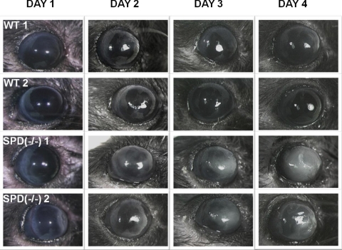
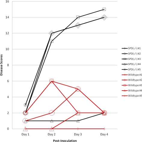
Methods: Murine corneas were blotted with tissue paper before inoculation with green fluorescent protein-expressing P. aeruginosa. The impact of blotting on epithelial integrity was evaluated by susceptibility to fluorescein staining and histology. Using fluorescence imaging, blotted corneas were compared to nonblotted corneas for susceptibility to bacterial binding and epithelial penetration after 5 hours or were monitored for disease development. In some experiments, inoculation was performed ex vivo to exclude tear fluid or corneas were pretreated with EGTA to disrupt Ca(2+)-dependent factors. The role of surfactant protein D (SP-D), which inhibits P. aeruginosa cell invasion in vitro, was examined using knockout mice.

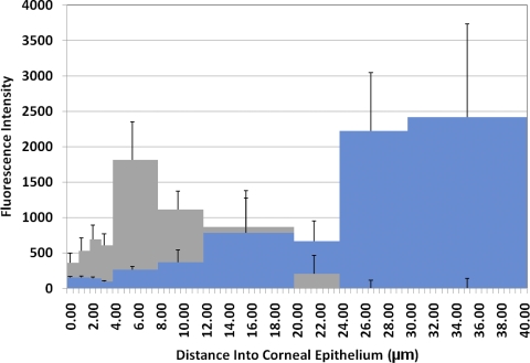
Results: Blotting enabled fluorescein penetration through the epithelium into the underlying stroma without obvious disruption to corneal morphology. Although blotting enabled bacterial binding to the otherwise adhesion-resistant epithelial surface, adherent bacteria did not penetrate the surface or initiate pathology. In contrast, bacteria penetrated blotted corneas after EGTA treatment and in SP-D knockouts. Visible disease occurred and progressed only in aged, blotted, and EGTA-treated, SP-D knockout mice.

Conclusions: Neither fluorescein staining nor bacterial adhesion necessarily predict or enable corneal susceptibility to bacterial penetration or disease. Corneal epithelial defenses limiting traversal by adherent bacteria include EGTA-sensitive factors and SP-D. Understanding mechanisms modulating epithelial traversal by microbes could improve our understanding of susceptibility to infection and may indicate new strategies for preventing disease.


Figures

Figure 1.
Figure 1.
Impact of tissue paper blotting on corneal susceptibility to fluorescein staining and infection. Fluorescein was added to wild-type (C57BL/6) corneas immediately after the blotting procedure. Blotted corneas showed extensive fluorescein staining (A). Confocal microscopy z-stack of blotted and fluorescein-stained mouse cornea showed fluorescein penetration of the corneal epithelium and stroma to a depth of approximately 100 μM (B). Other blotted corneas were immediately inoculated with GFP-expressing P. aeruginosa strain PAO1. Blotting did not enable susceptibility to infection at 24 hours after infection (C).
Figure 2.
Figure 2.
(A) Transverse section through rinsed and tissue paper–blotted mouse cornea (C57BL/6). Toluidine-blue staining illustrated that the corneal epithelium remained multilayered and morphologically indistinguishable from a normal healthy cornea (B) after these treatments.
Figure 3.
Figure 3.
(A, B) Bacterial distribution within wild-type (C57BL/6) cornea inoculated with GFP-expressing P. aeruginosa strain PAO1for 5 hours in vivo, as assessed using fluorescence imaging. Healthy corneas were clear of inoculated bacteria (A). Blotted corneas showed bacterial adherence to the superficial surface (arrow), but bacteria were not detected below the surface within deeper layers of the corneal epithelium (B). (C, D) Impact of ex vivo inoculation on susceptibility of the blotted cornea to P. aeruginosa penetration. The impact of excluding tear fluid was examined by comparing in vivo inoculation to inoculation of excised eyeballs in vitro. Corneas of wild-type (C57BL/6) mice were tissue paper blotted, then inoculated with GFP-expressing P. aeruginosa strain PAO1 (green) either in vivo (C) or after excision ex vivo (D) for 5 hours. In both situations, bacteria adhered to the corneal epithelium (arrows) but did not traverse. Scale bar, 10 μm. Green: bacteria (GFP); blue: cell nuclei (DAPI); red: laminin (rhodamine).
Figure 4.
Figure 4.
(AD) Impact of EGTA pretreatment on the susceptibility to P. aeruginosa traversal. Blotted murine (C57BL/6) eyeballs were pretreated ex vivo for 1 hour with PBS (A) or with 100 mM EGTA in PBS (BD) before incubation with GFP-expressing bacteria. After 5 hours, significant numbers of bacteria (arrows) traversed the corneal epithelia of EGTA-pretreated, but not PBS control, eyes. Scale bar, 10 μm. Green: bacteria (GFP); blue: cell nuclei (DAPI); red: laminin (rhodamine). (E, F) Immunofluorescence (ZO-1 labeling) of cryosectioned mouse corneas. Enucleated, blotted, and PBS rinsed murine (Black Swiss) eyeballs were pretreated ex vivo for 1 hour with PBS or with100 mM EGTA in PBS. Blotted, PBS-treated corneas showed ZO-1 labeling throughout the epithelium (E), which was diffused in the blotted, EGTA-pretreated corneas (F). Scale bar, 10 μm. Blue: nuclei (DAPI); red: ZO-1 (rhodamine).
Figure 5.
Figure 5.
Quantification of GFP-labeled P. aeruginosa penetration of the blotted murine cornea (Black Swiss) ex vivo with and without EGTA pretreatment. After euthanatization, eyes were enucleated, tissue paper blotted, and PBS rinsed before pretreatment ex vivo for 1 hour with PBS or EGTA (100 mM in PBS). After removal of the EGTA solution and a PBS wash, eyes were incubated with GFP-expressing P. aeruginosa (200 μL of a concentrated bacterial suspension approximately 1011 cfu/mL). After 6 hours of exposure to bacteria, confocal microscopy showed greater bacterial traversal of the corneal epithelium in EGTA-pretreated eyes (blue) compared with PBS-treated controls (gray).
Figure 6.
Figure 6.
(AD) Impact of SP-D on epithelial susceptibility to P. aeruginosa traversal. Corneas of wild-type C57BL/6 (A) and SP-D−/− (BD) mice were tissue paper blotted before inoculation in vivo with GFP-expressing P. aeruginosa strain PAO1. Fluorescence microscopy showed that after 5 hours, bacteria had partially traversed the corneal epithelium of SP-D−/−, but not wild-type, mice. Scale bar, 10 μm. (green) Bacteria (GFP). Blue: cell nuclei (DAPI); red: laminin (rhodamine). (E) Quantification of GFP-labeled P. aeruginosa penetration of a blotted cornea ex vivo comparing wild-type (Black Swiss) and SP-D−/− mice. After euthanatization, eyes were enucleated, tissue paper blotted, and PBS rinsed before incubation with GFP-expressing P. aeruginosa (200 μL of a concentrated bacterial suspension approximately 1011 cfu/mL). After 8 hours of exposure to bacteria, confocal microscopy showed greater bacterial traversal of the corneal epithelium in SP-D−/− eyes (blue) compared with wild-type (gray).
Figure 7.
Figure 7.
Examples of corneal disease found in aged (35–36 weeks) wild-type (C57BL/6) mice and SP-D−/− mice 1 to 4 days after inoculation with P. aeruginosa strain PAO1 (approximately 109 cfu in 5 μL). Corneas were blotted and then treated with EGTA (100 mM in PBS) for 3 hours before inoculation. Two-millimeter cutouts from a soft contact lens were used to retain EGTA at the ocular surface for the 3-hour period. Disease in SP-D−/− mice became progressively more severe with time; in contrast, wild-type eyes with visible pathology at 24 hours showed little further progression or recovered.
Figure 8.
Figure 8.
Progression of corneal disease (individual disease scores) in aged mice (C57BL/6) compared with aged-matched SP-D−/− mice after inoculation with P. aeruginosa (approximately 109 cfu in 5 μL). Before inoculation, eyes were pretreated with EGTA (Fig. 7). Significant differences in disease scores were observed between wild-type and SP-D−/− groups on each day (t-tests: day 1, P = 0.016; day 2, P = 0.027; day 3, P = 0.045; day 4, P = 0.03). One mouse (SP-D−/− mouse 5) died after day 1.

Comment in

References

    1. Cheng KH, Leung SL, Hoekman HW, et al. Incidence of contact-lens-associated microbial keratitis and its related morbidity. Lancet. 1999;354:181–185 - PubMed
    1. Cruciani F, Cuozzo G, Di Pillo S, Cavallaro M. Predisposing factors, clinical and microbiological aspects of bacterial keratitis: a clinical study. Clin Ther. 2009;160:207–210 - PubMed
    1. Stapleton F, Keay L, Edwards K, et al. The incidence of contact lens-related microbial keratitis in Australia. Ophthalmology. 2008;115:1655–1662 - PubMed
    1. Mun JJ, Tam C, Kowbel D, et al. Clearance of Pseudomonas aeruginosa from a healthy ocular surface involves surfactant protein D and is compromised by bacterial elastase in a murine null-infection model. Infect Immun. 2009;77:2392–2398 - PMC - PubMed
    1. Hazlett LD. Corneal response to Pseudomonas aeruginosa infection. Prog Retin Eye Res. 2004;23:1–30 - PubMed

Publication types

MeSH terms

Substances