. 2011 Feb 16;52(2):959-68.
doi: 10.1167/iovs.10-6207.

The oral iron chelator deferiprone protects against iron overload-induced retinal degeneration

Affiliations

The oral iron chelator deferiprone protects against iron overload-induced retinal degeneration

Majda Hadziahmetovic et al. Invest Ophthalmol Vis Sci. .

Abstract

Purpose: Iron-induced oxidative stress may exacerbate age-related macular degeneration (AMD). Ceruloplasmin/Hephaestin double-knockout (DKO) mice with age-dependent retinal iron accumulation and some features of AMD were used to test retinal protection by the oral iron chelator deferiprone (DFP).

Methods: Cultured retinal pigment epithelial (ARPE-19) cells and mice were treated with DFP. Transferrin receptor mRNA (Tfrc), an indicator of iron levels, was quantified by qPCR. In mice, retinal oxidative stress was assessed by mass spectrometry, and degeneration by histology and electroretinography.

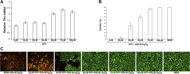
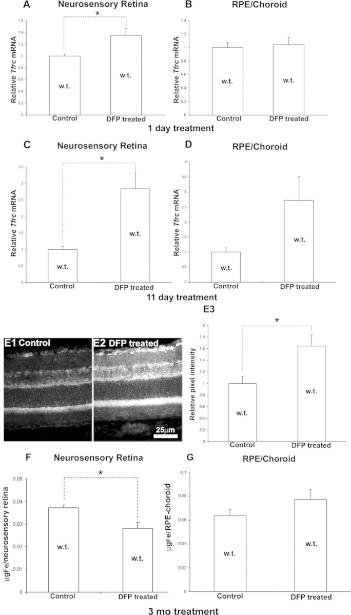
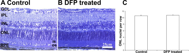
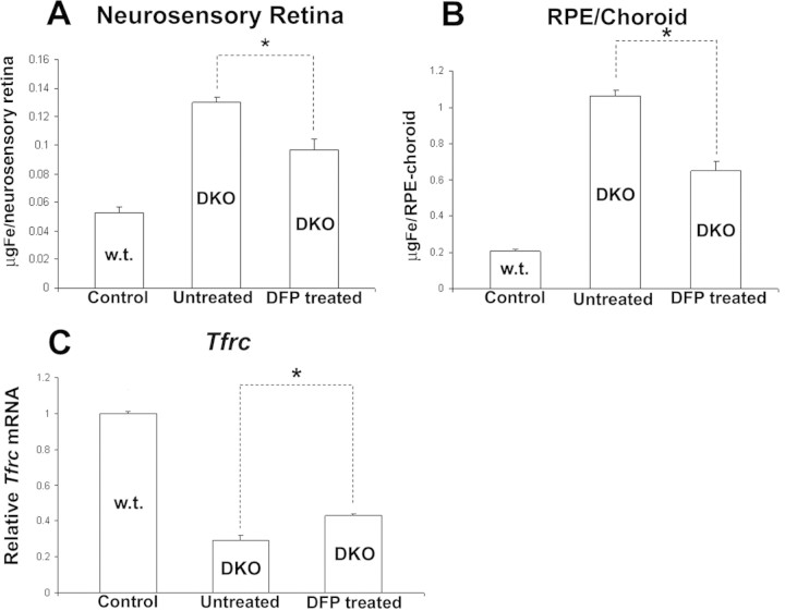
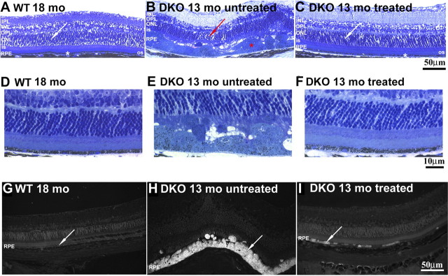
Results: DFP at 60 μM decreased labile iron in ARPE-19 cells, increasing Tfrc and protecting 70% of cells against a lethal dose of H(2)O(2). DFP 1 mg/mL in drinking water increased retinal Tfrc mRNA 2.7-fold after 11 days and also increased transferrin receptor protein. In DKOs, DFP over 8 months decreased retinal iron levels to 72% of untreated mice, diminished retinal oxidative stress to 70% of the untreated level, and markedly ameliorated retinal degeneration. DFP was not retina toxic in wild-type (WT) or DKO mice, as assessed by histology and electroretinography.

Conclusions: Oral DFP was not toxic to the mouse retina. It diminished retinal iron levels and oxidative stress and protected DKO mice against iron overload-induced retinal degeneration. Further testing of DFP for retinal disease involving oxidative stress is warranted.


Figures

Figure 1.
Figure 1.
DFP treatment decreased labile iron and protected ARPE19 cells from H2O2-induced cell death. (A) Relative Tfrc mRNA levels as determined by qPCR in ARPE-19 cells after 15 hours of treatment with different concentrations of DFP, as indicated. (B, C) ARPE-19 cells were treated for 15 hours with MEM (100% viability control), 200 μM H2O2 alone, or 200 μM H2O2 with different concentrations of DFP, as indicated. (B) Percentage of cell viability as determined by LDH assay. DFP protected the ARPE-19 cells against H2O2-induced cell death. (C) Fluorescence photomicrographs of ARPE-19 cells labeled in a fluorescent cell viability assay. Red: dead cells as detected by ethidium bromide homodimer fluorescence. Green: live cells as detected by calcein fluorescence. Error bars ± SEM.
Figure 2.
Figure 2.
DFP treatment in WT mice chelated labile iron, increasing retinal Tfrc mRNA and protein levels. Wild-type mice had significantly increased Tfrc mRNA levels, as measured by qPCR in the neural retina after DFP treatment for 1 day (A; n = 3 mice per group, 6 months old) and 11 days (C; n = 4 mice per group, 6 months old). The difference in Tfrc expression is not statistically significant in the RPE/choroid in both groups (B, D). Representative fluorescence retinal photomicrographs from 11-day untreated control (E1) and DFP-treated (E2) WT mice. The DFP-treated mice showed increased anti-TfR immunoreactivity throughout the retina. Scale bar, 25 μm. Immunoreactivity was quantified by measuring the mean pixel intensity within the RPE and neural retinas (n = 3 mice per group, 6 months old) and is shown as the mean (E3). Three months of treatment with DFP significantly reduced total iron in the neural retina (F), but not in the RPE/choroid (G). *Significant difference (P < 0.05). Error bars ± SEM.
Figure 3.
Figure 3.
Normal retinal morphology after DFP treatment. Representative bright-field photomicrographs from WT DFP-treated (B) and untreated (A) 9-month-old mice (n = 3 each). After 3 months of treatment, the retinas showed no changes in morphology or in the number of ONL nuclei compared with the controls (C, P > 0.05). OS, photoreceptor outer segment; IS, photoreceptor inner segment; ONL, outer nuclear layer; OPL, outer plexiform layer; INL, inner nuclear layer; IPL, inner plexiform layer; GCL, ganglion cell layer. Scale bar, 25 μm.
Figure 4.
Figure 4.
DFP treatment decreased total nonheme and labile iron levels in DKO mice. Shown is total nonheme iron levels in 9-month-old DKO mice treated with DFP for 6 months relative to age-matched, untreated controls. Treated mice had significantly reduced total iron levels in the neural retina (A) and RPE/choroid (B). After 6 months of treatment with DFP, labile iron was significantly reduced in 8-month-old treated DKO mice as assessed by TfR mRNA upregulation (C). *Significant difference (P < 0.05). n = 3 mice per group. Error bars ± SEM.
Figure 5.
Figure 5.
Eight months of DFP treatment protected against iron-induced retinal degeneration and reduced the accumulation of lipofuscin-like material. Bright-field photomicrographs of plastic sections showed that 13-month-old untreated DKO mice (B, n = 3) had massively hypertrophic RPE cells (red asterisk) and focal thinning of the ONL (red arrow). In contrast, 13- to 14-month-old DKO mice treated for 8 months with DFP had normal morphology in most of the retina (C, white asterisk and white arrow; n = 4). (DF) Bright-field photomicrographs of plastic sections showing the same retinal changes under higher magnification. Fluorescence photomicrographs (Cy2 filter set) of plastic sections show a decrease in the number of autofluorescent RPE cells in DFP-treated DKO mice (I, arrow; n = 4), while the age-matched, untreated DKOs have autofluorescent RPE cells (H, arrow; n = 3) throughout most of the retina. All comparisons were made relative to 18-month WT mice that showed normal retinal architecture and absence of RPE autofluorescence (A, G, arrow, n = 3). RPE, retinal pigment epithelium; OS, photoreceptor outer segment; IS, photoreceptor inner segment; ONL, outer nuclear layer; OPL, outer plexiform layer; INL, inner nuclear layer; IPL, inner plexiform layer; GCL, ganglion cell layer. Scale bar: (AC, GI) 50 μm; (DF) 10 μm.
Figure 6.
Figure 6.
DFP treatment decreases retinal markers of oxidative stress. Graph showing retinal isoprostane F2α-VI levels in the retinas of 9-month-old DKO mice treated with DFP for 5 months relative to untreated, age-matched DKO and WT mice (A, n = 3 mice per group). DFP treatment significantly reduced Epo mRNA levels, as measured by qPCR, in the retinas of DKO mice treated for 6 months with DFP relative to untreated 9-month-old DKOs (B, n = 3 mice per group). DFP did not change Vegfa mRNA levels in either neurosensory retina (C) or RPE/choroid (D), but it significantly reduced Cd68 mRNA levels in neurosensory retinas of treated DKO relative to untreated DKO controls (E) and showed a trend toward C3 mRNA level reduction in the RPE/choroid (F). *Significant difference (P < 0.05). Error bars ± SEM.

Similar articles

Cited by

References

    1. Hahn P, Milam AH, Dunaief JL. Maculas affected by age-related macular degeneration contain increased chelatable iron in the retinal pigment epithelium and Bruch's membrane. Arch Ophthalmol. 2003;121:1099–1105 - PubMed
    1. Kontoghiorghes GJ, Efstathiou A, Kleanthous M, Michaelides Y, Kolnagou A. Risk/benefit assessment, advantages over other drugs and targeting methods in the use of deferiprone as a pharmaceutical antioxidant in iron loading and non iron loading conditions. Hemoglobin. 2009;33:386–397 - PubMed
    1. Snyder AM, Connor JR. Iron, the substantia nigra and related neurological disorders. Biochim Biophys Acta. 2009;1790:606–614 - PubMed
    1. Sullivan JL. Iron in arterial plaque: modifiable risk factor for atherosclerosis. Biochim Biophys Acta. 2009;1790:718–723 - PubMed
    1. Zacharski LR, Chow BK, Howes PS, et al. Decreased cancer risk after iron reduction in patients with peripheral arterial disease: results from a randomized trial. J Natl Cancer Inst. 2008;100:996–1002 - PubMed

Publication types

MeSH terms