

cIAP1 and TAK1 protect cells from TNF-induced necrosis by preventing RIP1/RIP3-dependent reactive oxygen species production
- PMID: 21052097
- PMCID: PMC3131911
- DOI: 10.1038/cdd.2010.138
cIAP1 and TAK1 protect cells from TNF-induced necrosis by preventing RIP1/RIP3-dependent reactive oxygen species production
Abstract
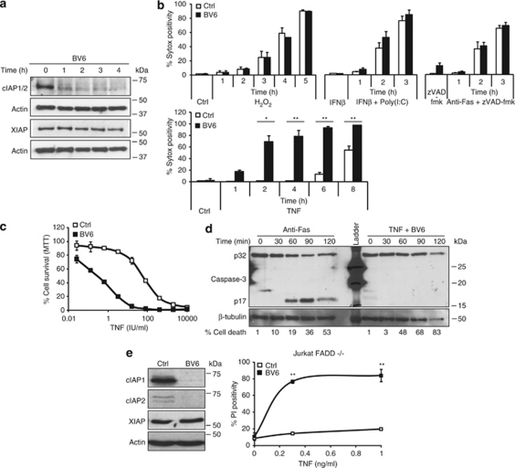
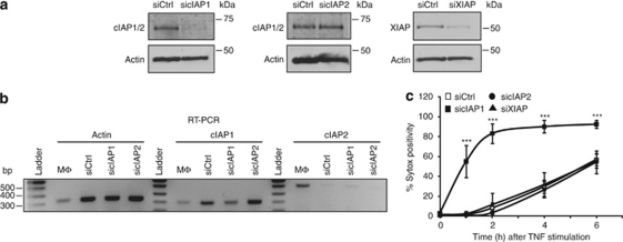
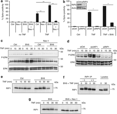
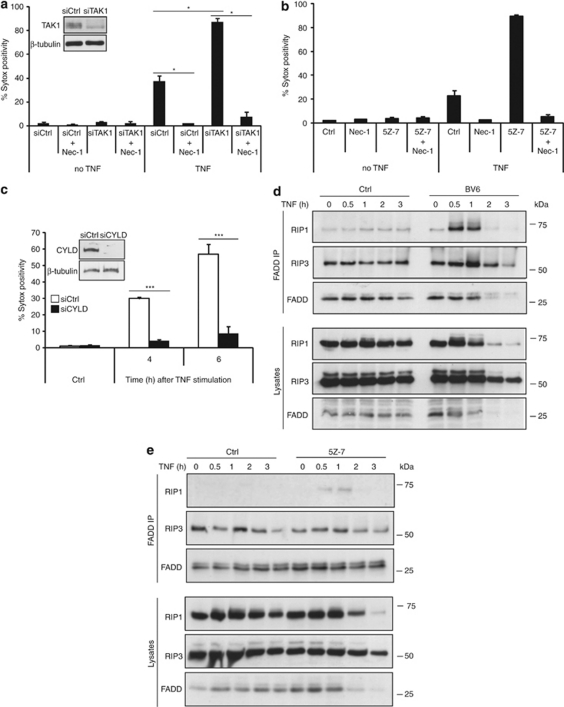
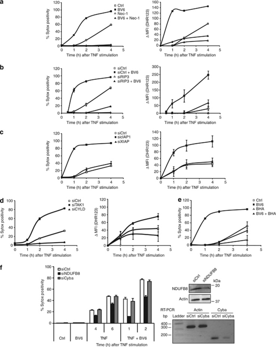
Three members of the IAP family (X-linked inhibitor of apoptosis (XIAP), cellular inhibitor of apoptosis proteins-1/-2 (cIAP1 and cIAP2)) are potent suppressors of apoptosis. Recent studies have shown that cIAP1 and cIAP2, unlike XIAP, are not direct caspase inhibitors, but block apoptosis by functioning as E3 ligases for effector caspases and receptor-interacting protein 1 (RIP1). cIAP-mediated polyubiquitination of RIP1 allows it to bind to the pro-survival kinase transforming growth factor-β-activated kinase 1 (TAK1) which prevents it from activating caspase-8-dependent death, a process reverted by the de-ubiquitinase CYLD. RIP1 is also a regulator of necrosis, a caspase-independent type of cell death. Here, we show that cells depleted of the IAPs by treatment with the IAP antagonist BV6 are greatly sensitized to tumor necrosis factor (TNF)-induced necrosis, but not to necrotic death induced by anti-Fas, poly(I:C) oxidative stress. Specific targeting of the IAPs by RNAi revealed that repression of cIAP1 is responsible for the sensitization. Similarly, lowering TAK1 levels or inhibiting its kinase activity sensitized cells to TNF-induced necrosis, whereas repressing CYLD had the opposite effect. We show that this sensitization to death is accompanied by enhanced RIP1 kinase activity, increased recruitment of RIP1 to Fas-associated via death domain and RIP3 (which allows necrosome formation), and elevated RIP1 kinase-dependent accumulation of reactive oxygen species (ROS). In conclusion, our data indicate that cIAP1 and TAK1 protect cells from TNF-induced necrosis by preventing RIP1/RIP3-dependent ROS production.
© 2011 Macmillan Publishers Limited
Figures
Similar articles
-
CYLD deubiquitinates RIP1 in the TNFα-induced necrosome to facilitate kinase activation and programmed necrosis.PLoS One. 2013 Oct 2;8(10):e76841. doi: 10.1371/journal.pone.0076841. eCollection 2013. PLoS One. 2013. PMID: 24098568 Free PMC article.
-
cIAP1 and cIAP2 limit macrophage necroptosis by inhibiting Rip1 and Rip3 activation.Cell Death Differ. 2012 Nov;19(11):1791-801. doi: 10.1038/cdd.2012.59. Epub 2012 May 11. Cell Death Differ. 2012. PMID: 22576661 Free PMC article.
-
Cellular IAP proteins and LUBAC differentially regulate necrosome-associated RIP1 ubiquitination.Cell Death Dis. 2015 Jun 25;6(6):e1800. doi: 10.1038/cddis.2015.158. Cell Death Dis. 2015. PMID: 26111062 Free PMC article.
-
The role of the kinases RIP1 and RIP3 in TNF-induced necrosis.Sci Signal. 2010 Mar 30;3(115):re4. doi: 10.1126/scisignal.3115re4. Sci Signal. 2010. PMID: 20354226 Review.
-
RIP1 post-translational modifications.Biochem J. 2022 May 13;479(9):929-951. doi: 10.1042/BCJ20210725. Biochem J. 2022. PMID: 35522161 Review.
Cited by
-
GRIM-19-mediated translocation of STAT3 to mitochondria is necessary for TNF-induced necroptosis.J Cell Sci. 2012 Jun 15;125(Pt 12):2995-3003. doi: 10.1242/jcs.103093. Epub 2012 Mar 5. J Cell Sci. 2012. Retraction in: J Cell Sci. 2016 Jul 1;129(13):2686. doi: 10.1242/jcs.193615. PMID: 22393233 Free PMC article. Retracted.
-
CYLD Proteolysis Protects Macrophages from TNF-Mediated Auto-necroptosis Induced by LPS and Licensed by Type I IFN.Cell Rep. 2016 Jun 14;15(11):2449-61. doi: 10.1016/j.celrep.2016.05.032. Epub 2016 Jun 2. Cell Rep. 2016. PMID: 27264187 Free PMC article.
-
Programmed necrosis and autophagy in immune function.Immunol Rev. 2012 Sep;249(1):205-17. doi: 10.1111/j.1600-065X.2012.01147.x. Immunol Rev. 2012. PMID: 22889224 Free PMC article. Review.
-
Reactive oxygen species regulate Smac mimetic/TNFα-induced necroptotic signaling and cell death.Oncogene. 2015 Nov 19;34(47):5796-806. doi: 10.1038/onc.2015.35. Epub 2015 Apr 6. Oncogene. 2015. PMID: 25867066
-
Tag7 (PGLYRP1) in Complex with Hsp70 Induces Alternative Cytotoxic Processes in Tumor Cells via TNFR1 Receptor.J Biol Chem. 2015 Aug 28;290(35):21724-31. doi: 10.1074/jbc.M115.639732. Epub 2015 Jul 16. J Biol Chem. 2015. PMID: 26183779 Free PMC article.
References
-
- Vandenabeele P, Galluzzi L, Vanden Berghe T, Kroemer G. Molecular mechanisms of necroptosis: an ordered cellular explosion. Nat Rev Mol Cell Biol. 2010;11:700–714. - PubMed
-
- Degterev A, Huang Z, Boyce M, Li Y, Jagtap P, Mizushima N, et al. Chemical inhibitor of nonapoptotic cell death with therapeutic potential for ischemic brain injury. Nat Chem Biol. 2005;1:112–119. - PubMed
-
- Vanden Berghe T, Vanlangenakker N, Parthoens E, Deckers W, Devos M, Festjens N, et al. Necroptosis, necrosis and secondary necrosis converge on similar cellular disintegration features. Cell Death Differ. 2010;17:922–930. - PubMed
Publication types
MeSH terms
Substances
LinkOut - more resources
Full Text Sources
Other Literature Sources
Molecular Biology Databases
Research Materials
Miscellaneous
