

A case of chorioretinal coloboma in a patient with achondroplasia
- PMID: 21052511
- PMCID: PMC2955274
- DOI: 10.3341/kjo.2010.24.5.302
A case of chorioretinal coloboma in a patient with achondroplasia
Abstract
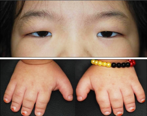
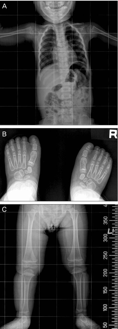
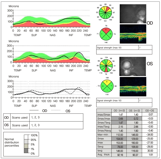
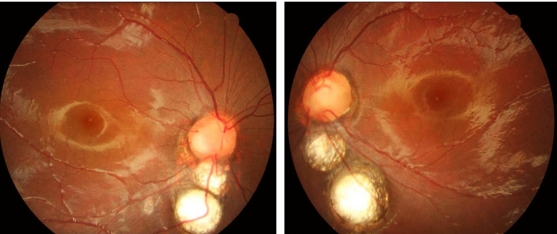
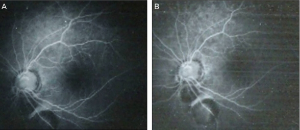
Achondroplasia is a congenital disorder resulting from a specific disturbance in endochondral bone formation. The ophthalmic features reportedly associated with achondroplasia are telecanthus, exotropia, inferior oblique overaction, angle anomalies and cone-rod dystrophy. This is first report of chorioretinal coloboma in achondroplasia. An 8-year-old female was diagnosed with a developmental delay, known as achondroplasia, seven months after birth. Upon her initial visit, visual acuity was 0.3 in both eyes. The patient had telecanthus but normal ocular motility. Findings were normal upon anterior segment examination. Fundus examination of both eyes revealed about 1,500 µm sized chorioretinal coloboma inferior to the optic nerve head. Upon fluorescent angiography, there was chorioretinal coloboma without any other lesions. Afterward, there was no change in the fundus lesion, and best corrected visual acuity was 0.6 in both eyes. Chorioretinal coloboma is associated with choroidal and retinal detachment. As chorioretinal coloboma and achondroplasia are developmental disorders in the embryonic stage, early detection and regular ophthalmologic examination would be essential in patients with achondroplasia.
Keywords: Achondroplasia; Chorioretinal coloboma.
Conflict of interest statement
No potential conflict of interest relevant to this article was reported.
Figures
Similar articles
-
Achondroplasia and Macular Coloboma.Middle East Afr J Ophthalmol. 2015 Oct-Dec;22(4):522-4. doi: 10.4103/0974-9233.167819. Middle East Afr J Ophthalmol. 2015. PMID: 26692730 Free PMC article.
-
SOLITARY RETINAL CAPILLARY HEMANGIOMA IN A PATIENT WITH BILATERAL CHORIORETINAL COLOBOMA.Retin Cases Brief Rep. 2019 Fall;13(4):320-323. doi: 10.1097/ICB.0000000000000586. Retin Cases Brief Rep. 2019. PMID: 28358746 Free PMC article.
-
Myriad of congenital excavated optic disc anomalies in achondroplasia.BMJ Case Rep. 2024 Nov 20;17(11):e261738. doi: 10.1136/bcr-2024-261738. BMJ Case Rep. 2024. PMID: 39572068
-
Bardet-Biedl syndrome with chorioretinal coloboma: a case series and review of literature.Ophthalmic Genet. 2024 Dec;45(6):616-622. doi: 10.1080/13816810.2024.2411257. Epub 2024 Oct 15. Ophthalmic Genet. 2024. PMID: 39402987 Review.
-
[Chorioretinal coloboma and neovascular membrane].Bull Soc Ophtalmol Fr. 1990 Jun-Jul;90(6-7):643-4. Bull Soc Ophtalmol Fr. 1990. PMID: 2225263 Review. French.
Cited by
-
Subluxated cataractous lens and high myopia: An uncommon association in an achondroplasia child.Oman J Ophthalmol. 2023 Oct 18;16(3):537-540. doi: 10.4103/ojo.ojo_42_23. eCollection 2023 Sep-Dec. Oman J Ophthalmol. 2023. PMID: 38059098 Free PMC article.
-
Achondroplasia associated with bilateral keratoconus.Case Rep Ophthalmol Med. 2012;2012:573045. doi: 10.1155/2012/573045. Epub 2012 Dec 4. Case Rep Ophthalmol Med. 2012. PMID: 23259098 Free PMC article.
-
Achondroplasia and Macular Coloboma.Middle East Afr J Ophthalmol. 2015 Oct-Dec;22(4):522-4. doi: 10.4103/0974-9233.167819. Middle East Afr J Ophthalmol. 2015. PMID: 26692730 Free PMC article.
References
-
- Vajo Z, Francomano CA, Wilkin DJ. The molecular and genetic basis of fibroblast growth factor receptor 3 disorders: the achondroplasia family of skeletal dysplasias, Muenke craniosynostosis, and Crouzon syndrome with acanthosis nigricans. Endocr Rev. 2000;21:23–39. - PubMed
-
- Richette P, Bardin T, Stheneur C. Achondroplasia: from genotype to phenotype. Joint Bone Spine. 2008;75:125–130. - PubMed
-
- Maroteaux P. Osteochondrodysplasie. In: Maroteaux P, editor. Les maladies osseuses de lénfant. 3rd ed. Paris: Medicine-Sciences Flammarion; 1998. pp. 55–56.
-
- Rosenthal AR, Ryan SJ, Jr, Horowitz P. Ocular manifestations of dwarfism. Trans Am Acad Ophthalmol Otolaryngol. 1972;76:1500–1518. - PubMed
-
- DeRespinis PA, Caputo AR, Wagner RS, Guo S. Duane's retraction syndrome. Surv Ophthalmol. 1993;38:257–288. - PubMed
Publication types
MeSH terms
LinkOut - more resources
Full Text Sources
Research Materials
