Estrogen antagonist and development of macular hole
- PMID: 21052512
- PMCID: PMC2955275
- DOI: 10.3341/kjo.2010.24.5.306
Estrogen antagonist and development of macular hole
Abstract
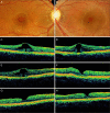
To describe the clinical and optical coherence tomography (OCT) features of a macular hole (MH) or its precursor lesion in patients treated with systemic antiestrogen agents. We reviewed the medical history of the patient, ophthalmic examination, and both fundus and OCT findings. Three female patients receiving antiestrogen therapy sought treatment for visual disturbance. All of the patients showed foveal cystic changes with outer retinal defect upon OCT. Visual improvement was achieved through surgery for the treatment of MH in two patients. Antiestrogen therapy may result in MH or its precursor lesion, in addition to perifoveal refractile deposits. OCT examination would be helpful for early detection in such cases.
Keywords: Antiestrogen agent; Foveal cyst; Retinal perforations; Tamoxifen; Toremifen.
Conflict of interest statement
No potential conflict of interest relevant to this article was reported.
Figures


References
-
- Bourla DH, Sarraf D, Schwartz SD. Peripheral retinopathy and maculopathy in high-dose tamoxifen therapy. Am J Ophthalmol. 2007;144:126–128. - PubMed
-
- Gualino V, Cohen SY, Delyfer MN, et al. Optical coherence tomography findings in tamoxifen retinopathy. Am J Ophthalmol. 2005;140:757–758. - PubMed
-
- Cronin BG, Lekich CK, Bourke RD. Tamoxifen therapy conveys increased risk of developing a macular hole. Int Ophthalmol. 2005;26:101–105. - PubMed
-
- Bernstein PS, DellaCroce JT. Diagnostic & therapeutic challenges. Tamoxifen toxicity. Retina. 2007;27:982–988. - PubMed
-
- Evans JR, Schwartz SD, McHugh JD, et al. Systemic risk factors for idiopathic macular holes: a case-control study. Eye (Lond) 1998;12(Pt 2):256–259. - PubMed
Publication types
MeSH terms
Substances
LinkOut - more resources
Full Text Sources

