. 2011:2011:391384.
doi: 10.1155/2011/391384. Epub 2010 Oct 31.

Mouse model resources for vision research

Affiliations

Mouse model resources for vision research

Jungyeon Won et al. J Ophthalmol. 2011.

Abstract

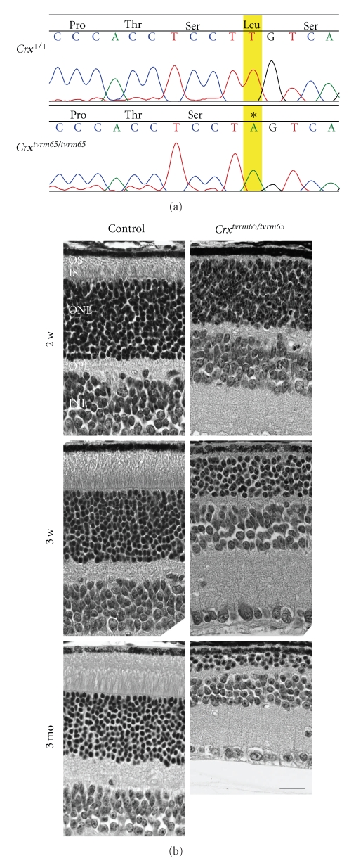
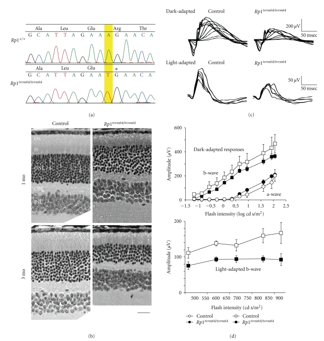
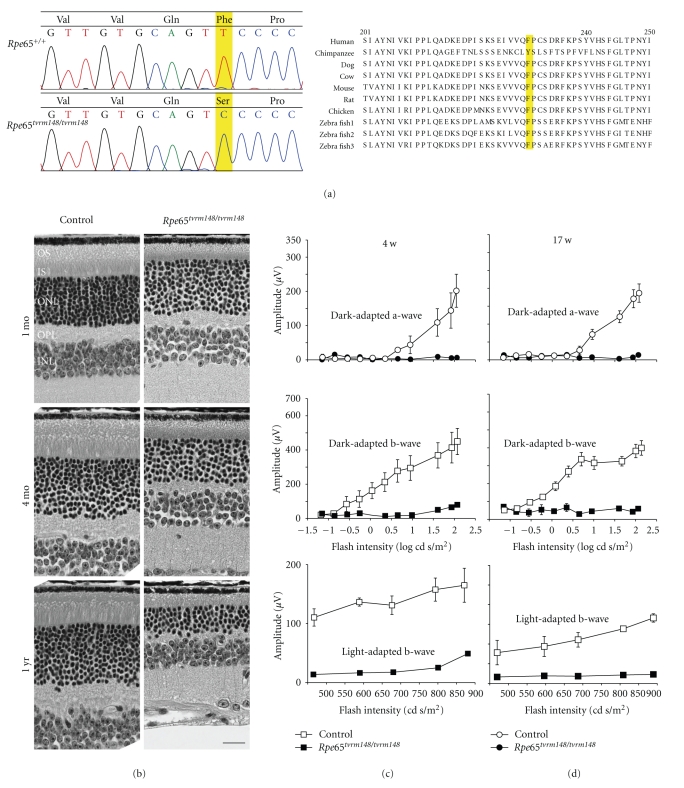
The need for mouse models, with their well-developed genetics and similarity to human physiology and anatomy, is clear and their central role in furthering our understanding of human disease is readily apparent in the literature. Mice carrying mutations that alter developmental pathways or cellular function provide model systems for analyzing defects in comparable human disorders and for testing therapeutic strategies. Mutant mice also provide reproducible, experimental systems for elucidating pathways of normal development and function. Two programs, the Eye Mutant Resource and the Translational Vision Research Models, focused on providing such models to the vision research community are described herein. Over 100 mutant lines from the Eye Mutant Resource and 60 mutant lines from the Translational Vision Research Models have been developed. The ocular diseases of the mutant lines include a wide range of phenotypes, including cataracts, retinal dysplasia and degeneration, and abnormal blood vessel formation. The mutations in disease genes have been mapped and in some cases identified by direct sequencing. Here, we report 3 novel alleles of Crx(tvrm65), Rp1(tvrm64), and Rpe65(tvrm148) as successful examples of the TVRM program, that closely resemble previously reported knockout models.


Figures

Figure 1
Figure 1
Schematic representation of the mating scheme of dominant (G1) or recessive (G3) screens. Male mice were mutagenized (3 weekly doses, 80 mg/kg) and mated to WT females after 4 weeks. If any female was pregnant within 5 weeks, the mating was discarded. If, however, male mice impregnated a female after that, the resulting G1 males were crossed to their respective female counterparts, and the G2 progeny were backcrossed to the G1 fathers to generate G3 offspring.
Figure 2
Figure 2
The mouse model C r x tvrm65. (a) The mutation in homozygous C r x tvrm65 causes a premature termination at aa residue Leu277. The mutated nucleotide is highlighted (b). Histology of control and C r x tvrm65 mutant retina at P14, P21, and 3 months of age. OSs were absent at all ages in homozygous C r x tvrm65, and progressive thinning of IS, ONL, and OPL was observed. OSs: outer segments, ISs: inner segments, ONL: outer nuclear layer, OPL: outer plexiform layer, INL: inner nuclear layer. Scale bar = 20 μm.
Figure 3
Figure 3
The mouse model R p1tvrm64. (a) Direct sequencing of control and R p1tvrm64 homozygous mutant identified an A to T mutation, predicting early termination at Arg522. The position of the mutation is highlighted and an asterisk indicates the termination. (b) The retinal morphology of control and R p1tvrm64 mice was examined at 1 and 3 months of age (mo). OSs: outer segments, ISs: inner segments, ONL: outer nuclear layer, OPL: outer plexiform layer, INL: inner nuclear layer. Scale bar = 20 μm. (c) Electroretinogram of dark-adapted (scotopic) and light-adapted (photopic) control at 9 weeks of age and R p1tvrm64 at 4 weeks of age. (d) The amplitude of dark-adapted a and b-wave and light-adapted b-wave (±SEM, n = 3) of 4 weeks old R p1tvrm64 mice and age matched controls.
Figure 4
Figure 4
The R p e65tvrm148 mouse model. (a) Mutation analysis by direct sequencing revealed that the homozygous R p e65tvrm148 mouse harbored a missense mutation at aa residue 229, causing an amino acid change from Phe to Ser. The highlighted nucleotide indicates the mutation in the R p e65tvrm148 mouse (left). RPE65 protein is an evolutionarily conserved protein, and F229 is a nearly invariant residue from human to zebra fish (right). (b) Retinal morphology at 1 and 4 months and 1 year of age was analyzed by light microscopy. ONL thinning was progressive, and IS/OS was shorter than controls at all ages examined. OSs: outer segments, ISs: inner segments, ONL: outer nuclear layer, OPL: outer plexiform layer, INL: inner nuclear layer. Scale bar = 20 μm. (c, d) Physiological retinal function was analyzed by ERG at 4 weeks (c) and 17 weeks of age (d). The plotted amplitude was obtained at 9 weeks from control and R p e65tvrm148 mice (c) or at 17 weeks from control and from homozygous R p e65tvrm148 mice. N = 3.

Similar articles

Cited by

References

    1. Chang B, Hawes N, Davisson M, Heckenlively J. Mouse models of RP. In: Tombran-Tink J, Barnstable C, editors. Retinal Degenerations: Biology, Diagnostics, and Therapeutics. The Human Press; 2007. pp. 149–164.
    1. Hawes NL, Smith RS, Chang B, Davisson M, Heckenlively JR, John SW. Mouse fundus photography and angiography: a catalogue of normal and mutant phenotypes. Molecular Vision. 1999;5:p. 22. - PubMed
    1. Wang W-H, Millar JC, Pang I-H, Wax MB, Clark AF. Noninvasive measurement of rodent intraocular pressure with a rebound tonometer. Investigative Ophthalmology and Visual Science. 2005;46(12):4617–4621. - PubMed
    1. Nusslein-Volhard C, Wieschaus E. Mutations affecting segment number and polarity in Drosophila. Nature. 1980;287(5785):795–801. - PubMed
    1. Kuwabara PE. Worming your way through the genome. Trends in Genetics. 1997;13(11):455–460. - PubMed