

Influence of age and sex steroids on bone density and geometry in middle-aged and elderly European men
- PMID: 21052641
- PMCID: PMC3073040
- DOI: 10.1007/s00198-010-1437-5
Influence of age and sex steroids on bone density and geometry in middle-aged and elderly European men
Abstract
Summary: The influence of age and sex steroids on bone density and geometry of the radius was examined in two European Caucasian populations. Age-related change in bone density and geometry was observed. In older men, bioavailable oestradiol may play a role in the maintenance of cortical and trabecular bone mineral density (BMD).
Introduction: To examine the effect of age and sex steroids on bone density and geometry of the radius in two European Caucasian populations.
Methods: European Caucasian men aged 40-79 years were recruited from population registers in two centres: Manchester (UK) and Leuven (Belgium), for participation in the European Male Ageing Study. Total testosterone (T) and oestradiol (E(2)) were measured by mass spectrometry and the free and bioavailable fractions calculated. Peripheral quantitative computed tomography was used to scan the radius at distal (4%) and midshaft (50%) sites.
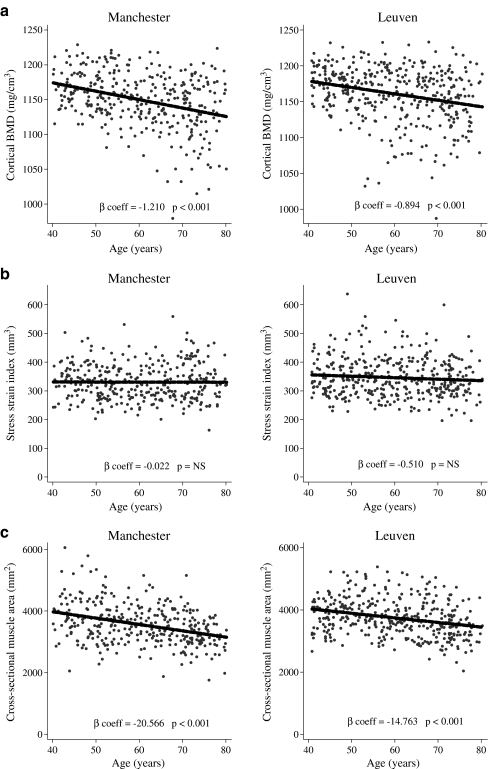
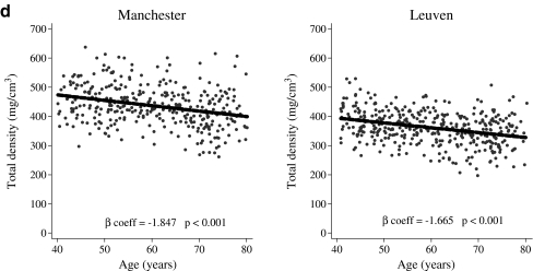
Results: Three hundred thirty-nine men from Manchester and 389 from Leuven, mean ages 60.2 and 60.0 years, respectively, participated. At the 50% radius site, there was a significant decrease with age in cortical BMD, bone mineral content (BMC), cortical thickness, and muscle area, whilst medullary area increased. At the 4% radius site, trabecular and total volumetric BMD declined with age. Increasing bioavailable E(2) (bioE(2)) was associated with increased cortical BMD (50% radius site) and trabecular BMD (4% radius site) in Leuven, but not Manchester, men. This effect was predominantly in those aged 60 years and over. In older Leuven men, bioavailable testosterone (Bio T) was linked with increased cortical BMC, muscle area and SSI (50% radius site) and total area (4% radius site).
Conclusions: There is age-related change in bone density and geometry at the midshaft radius in middle-aged and elderly European men. In older men bioE(2) may maintain cortical and trabecular BMD. BioT may influence bone health through associations with muscle mass and bone area.
Figures
Similar articles
-
Bone turnover predicts change in volumetric bone density and bone geometry at the radius in men.Osteoporos Int. 2017 Mar;28(3):935-944. doi: 10.1007/s00198-016-3816-z. Epub 2016 Nov 4. Osteoporos Int. 2017. PMID: 27815569 Free PMC article.
-
Role of sex steroids in the regulation of bone morphology in men. The MINOS study.Osteoporos Int. 2004 Nov;15(11):909-17. doi: 10.1007/s00198-004-1635-0. Epub 2004 Jul 3. Osteoporos Int. 2004. PMID: 15235765
-
Endogenous sex steroids and bone mineral density in older women and men: the Rancho Bernardo Study.J Bone Miner Res. 1997 Nov;12(11):1833-43. doi: 10.1359/jbmr.1997.12.11.1833. J Bone Miner Res. 1997. PMID: 9383688
-
Relationship of volumetric BMD and structural parameters at different skeletal sites to sex steroid levels in men.J Bone Miner Res. 2005 May;20(5):730-40. doi: 10.1359/JBMR.041228. Epub 2004 Dec 20. J Bone Miner Res. 2005. PMID: 15824845
-
Sex hormones and skeletal muscle weakness.Biogerontology. 2013 Jun;14(3):231-45. doi: 10.1007/s10522-013-9425-8. Epub 2013 May 1. Biogerontology. 2013. PMID: 23636830 Review.
Cited by
-
Androgens and estrogens in skeletal sexual dimorphism.Asian J Androl. 2014 Mar-Apr;16(2):213-22. doi: 10.4103/1008-682X.122356. Asian J Androl. 2014. PMID: 24385015 Free PMC article. Review.
-
The ESR1 (6q25) locus is associated with calcaneal ultrasound parameters and radial volumetric bone mineral density in European men.PLoS One. 2011;6(7):e22037. doi: 10.1371/journal.pone.0022037. Epub 2011 Jul 7. PLoS One. 2011. PMID: 21760950 Free PMC article.
-
Causality between Sex Hormones and Bone Mineral Density in Childhood: Age- and Tanner-Stage-Matched Sex Hormone Level May Be an Early Indicator of Pediatric Bone Fragility.Biomedicines. 2024 May 25;12(6):1173. doi: 10.3390/biomedicines12061173. Biomedicines. 2024. PMID: 38927380 Free PMC article.
-
Bone Mineral Density in the Noninstitutionalized Elderly: Influence of Sociodemographic and Anthropometric Factors.Curr Gerontol Geriatr Res. 2016;2016:4946593. doi: 10.1155/2016/4946593. Epub 2016 Apr 5. Curr Gerontol Geriatr Res. 2016. PMID: 27127504 Free PMC article.
-
Predicting ex vivo failure loads in human metatarsals using bone strength indices derived from volumetric quantitative computed tomography.J Biomech. 2013 Feb 22;46(4):745-50. doi: 10.1016/j.jbiomech.2012.11.019. Epub 2012 Dec 6. J Biomech. 2013. PMID: 23219276 Free PMC article.
References
-
- Burger H, de Laet CE, van Daele PL, Weel AE, Witteman JC, Hofman A, Pols HA. Risk factors for increased bone loss in an elderly population: the Rotterdam Study. Am J Epidemiol. 1998;147:871–879. - PubMed
Publication types
MeSH terms
Substances
Grants and funding
LinkOut - more resources
Full Text Sources
Medical
