Conserved cysteine residues within the attachment G glycoprotein of respiratory syncytial virus play a critical role in the enhancement of cytotoxic T-lymphocyte responses
- PMID: 21053062
- PMCID: PMC5454483
- DOI: 10.1007/s11262-010-0545-9
Conserved cysteine residues within the attachment G glycoprotein of respiratory syncytial virus play a critical role in the enhancement of cytotoxic T-lymphocyte responses
Abstract
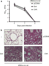
The cytotoxic T-lymphocyte (CTL) response plays an important role in the control of respiratory syncytial virus (RSV) replication and the establishment of a Th1-CD4+ T cell response against the virus. Despite lacking Major Histocompatibility Complex I (MHC I)-restricted epitopes, the attachment G glycoprotein of RSV enhances CTL activity toward other RSV antigens, and this effect depends on its conserved central region. Here, we report that RSV-G can also improve CTL activity toward antigens from unrelated pathogens such as influenza, and that a mutant form of RSV-G lacking four conserved cysteine residues at positions 173, 176, 182, and 186 fails to enhance CTL responses. Our results indicate that these conserved residues are essential for the wide-spectrum pro-CTL activity displayed by the protein.
Figures


Similar articles
-
The cysteine-rich region and secreted form of the attachment G glycoprotein of respiratory syncytial virus enhance the cytotoxic T-lymphocyte response despite lacking major histocompatibility complex class I-restricted epitopes.J Virol. 2006 Jun;80(12):5854-61. doi: 10.1128/JVI.02671-05. J Virol. 2006. PMID: 16731924 Free PMC article.
-
Combining DNA and protein vaccines for early life immunization against respiratory syncytial virus in mice.Eur J Immunol. 1999 Oct;29(10):3390-400. doi: 10.1002/(SICI)1521-4141(199910)29:10<3390::AID-IMMU3390>3.0.CO;2-A. Eur J Immunol. 1999. PMID: 10540351
-
Plasmid DNA encoding the respiratory syncytial virus G protein is a promising vaccine candidate.Virology. 2000 Mar 30;269(1):54-65. doi: 10.1006/viro.2000.0186. Virology. 2000. PMID: 10725198
-
The cytolytic activity of pulmonary CD8+ lymphocytes, induced by infection with a vaccinia virus recombinant expressing the M2 protein of respiratory syncytial virus (RSV), correlates with resistance to RSV infection in mice.J Virol. 1993 Feb;67(2):1044-9. doi: 10.1128/JVI.67.2.1044-1049.1993. J Virol. 1993. PMID: 8419638 Free PMC article.
-
Contribution of respiratory syncytial virus G antigenicity to vaccine-enhanced illness and the implications for severe disease during primary respiratory syncytial virus infection.Pediatr Infect Dis J. 2004 Jan;23(1 Suppl):S46-57. doi: 10.1097/01.inf.0000108192.94692.d2. Pediatr Infect Dis J. 2004. PMID: 14730270 Review.
Cited by
-
Respiratory syncytial virus G protein CX3C motif impairs human airway epithelial and immune cell responses.J Virol. 2013 Dec;87(24):13466-79. doi: 10.1128/JVI.01741-13. Epub 2013 Oct 2. J Virol. 2013. PMID: 24089561 Free PMC article.
-
Nanoparticle vaccines encompassing the respiratory syncytial virus (RSV) G protein CX3C chemokine motif induce robust immunity protecting from challenge and disease.PLoS One. 2013 Sep 10;8(9):e74905. doi: 10.1371/journal.pone.0074905. eCollection 2013. PLoS One. 2013. PMID: 24040360 Free PMC article.
-
Immunopathology of RSV: An Updated Review.Viruses. 2021 Dec 10;13(12):2478. doi: 10.3390/v13122478. Viruses. 2021. PMID: 34960746 Free PMC article. Review.
-
Persistence and continuous evolution of the human respiratory syncytial virus in northern Taiwan for two decades.Sci Rep. 2019 Mar 18;9(1):4704. doi: 10.1038/s41598-019-41332-9. Sci Rep. 2019. PMID: 30886248 Free PMC article.
-
Functional Analysis of the 60-Nucleotide Duplication in the Respiratory Syncytial Virus Buenos Aires Strain Attachment Glycoprotein.J Virol. 2015 Aug;89(16):8258-66. doi: 10.1128/JVI.01045-15. Epub 2015 May 27. J Virol. 2015. PMID: 26018171 Free PMC article.
References
Publication types
MeSH terms
Substances
Grants and funding
LinkOut - more resources
Full Text Sources
Research Materials

