

Odontogenic epithelium: immunolabeling of Ki-67, EGFR and survivin in pericoronal follicles, dentigerous cysts and keratocystic odontogenic tumors
- PMID: 21053110
- PMCID: PMC3037468
- DOI: 10.1007/s12105-010-0216-0
Odontogenic epithelium: immunolabeling of Ki-67, EGFR and survivin in pericoronal follicles, dentigerous cysts and keratocystic odontogenic tumors
Abstract
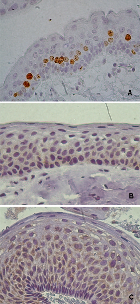
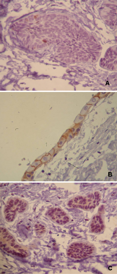
The aim of this study was to evaluate the biological profile of odontogenic epithelium by immunolabeling of epidermal growth factor receptor (EGFR), Ki-67 and survivin in keratocystic odontogenic tumors (KOT), dentigerous cysts (DC), and pericoronal follicles (PF). Immunohistochemical analysis was performed in 13 KOTs, 14 DCs and 9 PFs. Immunolabeling was analyzed in the basal and suprabasal layers of KOTs and DCs, and in the islands of odontogenic epithelium and/or reduced enamel epithelium of PFs. KOTs showed the highest proliferation rate among the three groups, mainly in suprabasal layers. EGFR immunolabeling was observed mainly in the cytoplasm in basal and suprabasal layers of KOTs and in the suprabasal layer of DCs. Immunolabeling in both membrane and cytoplasm was greater in PFs. In PFs, membrane-only staining was observed. Survivin immunolabeling showed a greater percentage of positive cells (scoring +++) in the suprabasal layer of KOTs. In DCs, both layers showed similar percentages of cells scoring +++; PFs showed the highest percentage of these cells. In KOTs, epithelial cells showed stimulus-independent neoplastic proliferative characteristics, suggesting the presence of a suprabasal proliferative compartment, maintained by inhibition of apoptosis. In DCs, the basal layer seemed to proliferate in response to stimulus. Although PFs showed low proliferative activity, the expression of EGFR indicates that some cells have a high capacity to respond to stimuli, which could probably explain the origin of odontogenic lesions.
Figures

Similar articles
-
The evaluation of Ki67, p53, MCM3 and PCNA immunoexpressions at the level of the dental follicle of impacted teeth, dentigerous cysts and keratocystic odontogenic tumors.Rom J Morphol Embryol. 2016;57(2):407-12. Rom J Morphol Embryol. 2016. PMID: 27516012
-
Cell proliferation and apoptosis in keratocystic odontogenic tumors.Med Oral Patol Oral Cir Bucal. 2008 Nov 1;13(11):E697-702. Med Oral Patol Oral Cir Bucal. 2008. PMID: 18978709
-
Immunohistochemical expression of p63, epidermal growth factor receptor (EGFR) and notch-1 in radicular cysts, dentigerous cysts and keratocystic odontogenic tumors.Braz Dent J. 2012;23(4):337-43. doi: 10.1590/s0103-64402012000400005. Braz Dent J. 2012. PMID: 23207846
-
Expression of Ki-67, p53 and p63 proteins in keratocyst odontogenic tumours: an immunohistochemical study.J Mol Histol. 2008 Jun;39(3):311-6. doi: 10.1007/s10735-008-9167-0. Epub 2008 Feb 7. J Mol Histol. 2008. PMID: 18256893
-
Odontogenic Cysts and Neoplasms.Surg Pathol Clin. 2017 Mar;10(1):177-222. doi: 10.1016/j.path.2016.10.006. Epub 2016 Dec 29. Surg Pathol Clin. 2017. PMID: 28153133 Review.
Cited by
-
Maspin, Syndecan-1, and Ki-67 in the Odontogenic Keratocyst: An Immunohistochemical Analysis.Int J Dent. 2020 Jul 14;2020:7041520. doi: 10.1155/2020/7041520. eCollection 2020. Int J Dent. 2020. PMID: 32733563 Free PMC article.
-
Computerized Evaluation of the Immunoexpression of Ki-67 Protein in Odontogenic Keratocyst and Dentigerous Cyst.Head Neck Pathol. 2020 Sep;14(3):598-605. doi: 10.1007/s12105-019-01077-3. Epub 2019 Sep 24. Head Neck Pathol. 2020. PMID: 31552621 Free PMC article.
-
Expression of human papillomavirus is correlated with Ki-67 and COX-2 expressions in keratocystic odontogenic tumor.Pathol Oncol Res. 2015 Jan;21(1):65-71. doi: 10.1007/s12253-014-9789-3. Epub 2014 May 15. Pathol Oncol Res. 2015. PMID: 24831259
-
A Comparison of Immunohistochemical Expression of Epidermal Growth Factor Receptor and Human Epidermal Growth Factor Receptor 2 in Dental Follicles with Different Radiographic Sizes.Iran J Med Sci. 2024 Aug 1;49(8):508-514. doi: 10.30476/IJMS.2023.98602.3121. eCollection 2024 Aug. Iran J Med Sci. 2024. PMID: 39205824 Free PMC article.
-
Immunohistochemical analysis of Ki-67 in dental follicle of asymptomatic impacted third molars.J Oral Maxillofac Pathol. 2014 May;18(2):189-93. doi: 10.4103/0973-029X.140737. J Oral Maxillofac Pathol. 2014. PMID: 25328297 Free PMC article.
References
-
- Baumgart SC, Lauxen IS, Sant’Ana Filho M, et al. Epidermal growth factor receptor distribution in pericoronal follicles: relationship with the origin of odontogenic cysts and tumors. Oral Surg Oral Med Oral Pathol Oral Radiol Endod. 2007;103:240–245. doi: 10.1016/j.tripleo.2005.11.009. - DOI - PubMed
-
- Edmatsu M, Kumanoto H, Ooya K, et al. Apoptosis-related factors in the epithelial components of pericoronal follicles and dentigerous cysts associated with impacted third molars of the mandible. Oral Surg Oral Med Oral Pathol Oral Radiol Endod. 2005;99:17–23. doi: 10.1016/j.tripleo.2004.04.016. - DOI - PubMed
MeSH terms
Substances
LinkOut - more resources
Full Text Sources
Research Materials
Miscellaneous
