. 2010 Sep-Oct;15(5):056001.
doi: 10.1117/1.3484262.

Imaging of normal and pathologic joint synovium using nonlinear optical microscopy as a potential diagnostic tool

Affiliations

Imaging of normal and pathologic joint synovium using nonlinear optical microscopy as a potential diagnostic tool

Nivedan Tiwari et al. J Biomed Opt. 2010 Sep-Oct.

Abstract

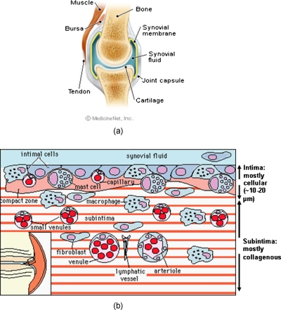
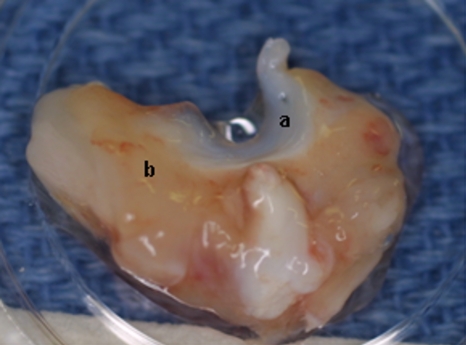
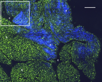
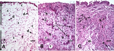
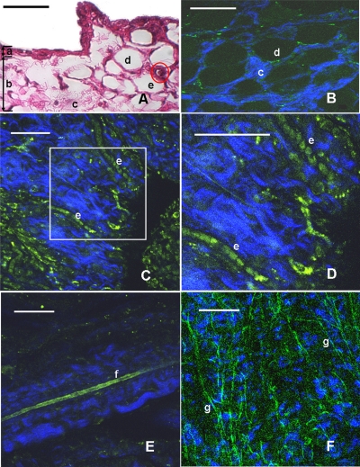
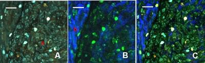
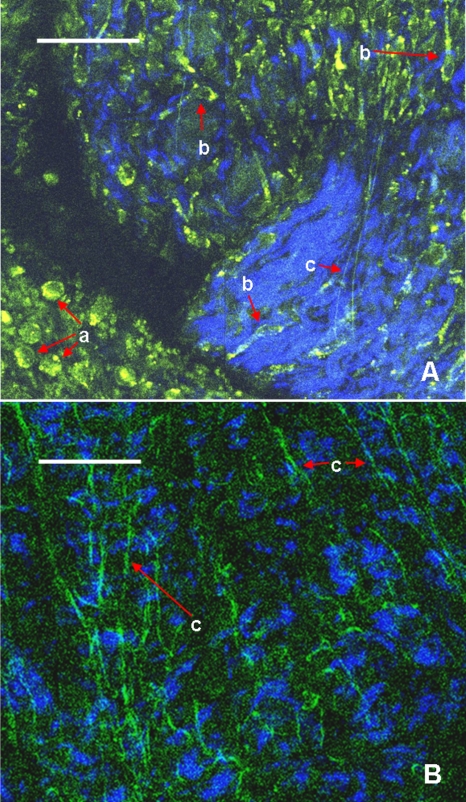
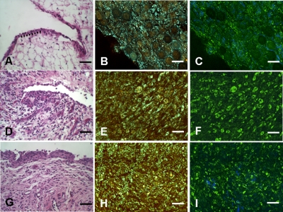
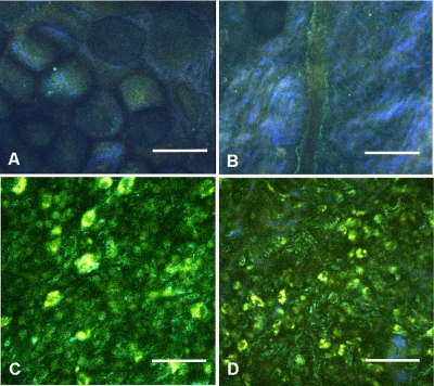
An estimated 1.3 million people in the United States suffer from rheumatoid arthritis (RA). RA causes profound changes in the synovial membrane of joints, and without early diagnosis and intervention, progresses to permanent alterations in joint structure and function. The purpose of this study is to determine if nonlinear optical microscopy (NLOM) can utilize the natural intrinsic fluorescence properties of tissue to generate images that would allow visualization of the structural and cellular composition of fresh, unfixed normal and pathologic synovial tissue. NLOM is performed on rabbit knee joint synovial samples using 730- and 800-nm excitation wavelengths. Less than 30 mW of excitation power delivered with a 40×, 0.8-NA water immersion objective is sufficient for the visualization of synovial structures to a maximum depth of 70 μm without tissue damage. NLOM imaging of normal and pathologic synovial tissue reveals the cellular structure, synoviocytes, adipocytes, collagen, vascular structures, and differential characteristics of inflammatory infiltrates without requiring tissue processing or staining. Further study to evaluate the ability of NLOM to assess the characteristics of pathologic synovial tissue and its potential role for the management of disease is warranted.


Figures

Figure 1
Figure 1
Schematic depictions of (a) diarthrodial joint structure and (b) the layered structure of synovium.
Figure 2
Figure 2
Schematic depiction of optical sectioning of thick synovial tissue using NLOM. Individual images obtained in tangential planes at specific depth intervals of the tissue (a) can be assembled into a 3-D tomographic stack of images (b) referred to as a Z stack.
Figure 3
Figure 3
(a) Meniscus with (b) synovium covered joint capsule attached to its lateral margin, as dissected from a rabbit knee joint. Retention of the meniscus allowed orientation and easy manipulation of the joint capsule tissue without disrupting the synovium.
Figure 4
Figure 4
A NLOM tiled image of the synovial surface layer constructed from sequentially acquired 225×225-μm images using 800-nm excitation. (a) The SHG emission from collagen is in blue, (b) TPF, in green, is mostly from intracellular sources, and (c) spaces between synovial folds lack any fluorescence signal. Scale bar is 100 μm. The box-enclosed area is further magnified in Fig. 8A to show cellular details. (Color online only.)
Figure 5
Figure 5
HE stained histological images of adipose synovium from (A) a knee joint of a normal rabbit, (B) 8 days postinoculation with S. aureus, and (C) 18 days postinoculation with LPS from E. coli. (A) In the cross section of normal adipose synovium, the intima (a) is seen as a densely populated cellular region that is one to three cells thick, while the subintima (b) is composed primarily of adipocytes (c) and small numbers of capillaries (d), small venules (e), and arterioles set in a randomly distributed matrix of loosely packed thin collagen fibrils (f). The sharp transition between the intima and subintima and the characteristic morphological details of each layer are easily observed. The cross section of adipose synovium from the S. aureus infected knee (B) demonstrates that a denuded intimal layer (g) and the adipocytes (c) of the thickened and edematous subintima have been largely replaced by an inflammatory cell infiltrate (h) consisting primarily of heterophils. Capillaries (i) are congested and their walls are thickened. Fibroblasts (j) are beginning to produce a delicate fibrillar matrix, while the meshwork of pre-existing collagen fibers is randomly displaced. The image of the inflammatory reaction that is present in the adipose synovium of the rabbit knee joint following LPS inoculation (C) demonstrates thickening of the intimal layer (a), and the normal population of subintimal adipocytes has been replaced by an inflammatory cell infiltrate (l) consisting primarily of lymphocytes relatively evenly distributed, along with fibroblasts in a collagenous matrix containing congested capillaries (i), arterioles (k), and venules (e). In some areas, edema (m) separates the matrix of collagen fibrils. Scale bars are 50 μm.
Figure 6
Figure 6
Important synovial structures as demonstrated by (A) HE sectioning and (B) through (F) NLOM imaging using 800-nm excitation. All images except for (D) are presented in the same scale for ease of comparison. The HE section shows the thin intimal layer (a) and the much thicker subintima (b). Collagen (c) appears as a widespread wavy pattern in (A) and is identified particularly well by its strong blue SHG signature in (B) through (F). Adipocytes (d) appear as empty spaces in the extracellular collagen matrix in the HE stained section (A) and as dark oval structures enclosed by the blue SHG signal of the collagen fibers in the NLOM image (B). Rouleaux formation of erythrocytes in undulating venuoles (e) set in a background meshwork of blue collagen fibrils of the subintima are seen in (C) and (D). (D) Image is a magnification of the area inside the box in (C), and demonstrates the visualization of individual erythrocytes. An arteriole (f) is seen in (A) and in (E), where the green colored TPF signal of elastin fibers outline the wall of a linearly oriented arteriole. Arteriole walls have elastin that provides a strong TPF signal, while the primarily collagen-containing venule walls produce only a SHG signal that highlights erythrocytes within the vascular lumen (e). Linearly directed elastin fibers appearing as green filaments (g) set in a meshwork of less-well-oriented blue collagen fibers are observed in synovial tissue along the insertion line of the adipose synovium with the meniscus (F). Scale bars are 50 μm for all images. (Color online only.)
Figure 7
Figure 7
Comparison of synovial images of the same location acquired using (A) 730-nm excitation and (B) 800-nm excitation at a pixel dwell time of 12.8 μsec. At this scan speed, cells (a) appear clearer with 730-nm excitation, while collagen (b) is most clearly distinguished when using 800-nm excitation. (C) An improved image was created by combining the TPF channels from (A) and SHG channel from (B). Scale bars are 20 μm.
Figure 8
Figure 8
Magnified NLOM images showing synovial cells and elastin fibers from (A) the box-enclosed area of Fig. 4, and (B) from Fig. 6F. Rounded synoviocytes, 10 to 20 μm in diameter (a), and elongated synoviocytes, 40 to 50 μm long (b), can be distinguished in (A). Long, thin elastin-like fibers (c) in (A) and (B) are observed to be morphologically distinct from the elongated synoviocytes in (A). Scale bars are 50 μm.
Figure 9
Figure 9
Images of rabbit synovial tissue, (A), (B), and (C) normal, (D), (E), and (F) 8 days postbacterial inoculation, and (G), (H), and (I) 18 days post-LPS intra-articular injection, demonstrating HE stained histology sections in (A), (D), and (G), NLOM using 730-nm excitation in (B), (E), and (H), and NLOM using 800-nm excitation in (C), (F), and (I). All NLOM images were acquired in a plane parallel to and at a depth of 15 μm from the specimen surface [dotted line, in (A)] and thus primarily image the inflammatory response within the intima. Histologic sections of the pathology specimens demonstrate a marked inflammatory response characterized by mixed inflammatory cells, macrophages, and fibroblasts in the septic specimens in (D) and a generalized and predominantly lymphocytic infiltration with pronounced intimal thickening observed in the synovium of the LPS-treated joints in (G). NLOM images of both pathology specimens in (E), (F), (H), and (I) reveal increased intracellular fluorescence consistent with the elevated level of metabolic activity of cells actively engaged in the inflammatory response. There is a conspicuous absence of collagen SHG observed in the septic tissue in (F), while collagen SHG (blue) continues to be observed in synovial tissue of the LPS-treated joints in (I). The larger size of the inflammatory cell types (heterophils and macrophages) with multilobulated nuclei and the elongated morphology of the fibroblasts is clearly evident in the NLOM images of the septic tissue in (E) and (F), while in lymphocytes that are morphologically smaller in size with an oval nucleus, minimal to no evidence of fibroblast activity and the continued observation of some collagen SHG characterize the pathology of the tissue from LPS-treated joints in (H) and (I). Scale bars are 50 μm. (Color online only.)
Figure 10
Figure 10
NLOM images of rabbit synovial tissue demonstrating the enhanced image quality achieved by combining images acquired by sequential 730-nm and then 800-nm excitation. The images illustrated were obtained from the subintima in a plane parallel to and at a depth of 45 μm from the surface of (A) and (B) normal, (C) 8 days postbacterial inoculation, and (D) 18 days post-LPS intra-articular injection specimens. At a depth of 45 μm, the imaging plane is below the intimal layer of the normal synovium and demonstrates subintimal tissue predominately consisting of (A) adipocytes or (B) collagen matrix with little other cellular content, while both the (C) septic and (D) LPS specimens demonstrate a substantial increase in cellular content consistent with the inflammatory processes. The septic tissue demonstrates large cells with multilobular nuclei characteristic of rabbit heterophils and macrophages, along with elongated cells characteristic of fibroblasts, while the cellular infiltrate observed in the LPS specimens are smaller cells with oval nuclei characteristic of an inflammatory response predominated by lymphocytes. SHG emission of collagen, viewed as blue fibrils in the NLOM images, is not observed in the subintimal tissue of the septic specimens in (C), while it continues to be observed, although in reduced amounts, in LPS specimens in (D). Scale bars are 50 μm. (Color online only.)

Similar articles

Cited by

References

    1. Helmick C. G., Felson D. T., Lawrence R. C., Gabriel S., Hirsch R., Kwoh C. K., Liang M. H., Kremers H. M., Mayes M. D., Merkel P. A., Pillemer S. R., Reveille J. D., and Stone J. H., “Estimates of the prevalence of arthritis and other rheumatic conditions in the United States. Part I,” Arthritis Rheum. ARHEAW 58, 15–25 (2008).10.1002/art.23177 - DOI - PubMed
    1. Lawrence R. C., Felson D. T., Helmick C. G., Arnold L. M., Choi H., Deyo R. A., Gabriel S., Hirsch R., Hochberg M. C., Hunder G. G., Jordan J. M., Katz J. N., Kremers H. M., and Wolfe F., “Estimates of the prevalence of arthritis and other rheumatic conditions in the United States. Part II,” Arthritis Rheum. ARHEAW 58, 26–35 (2008).10.1002/art.23176 - DOI - PMC - PubMed
    1. Gerlag D. M. and Tak P. P., “Synovial fluid analysis, synovial biopsy, and synovial pathology,” in Kelly’s Textbook of Rheumatology, Ruddy S., Harris E. D., and Sledge C. B., Eds., pp. 675–691, W. B. Saunders Co., Philadelphia, PA: (2001).
    1. Lee D. M., Kiener G. P., and Brenner M. B., “Synoviocytes,” in Kelly’s Textbook of Rheumatology, Ruddy S., Harris E. D., and Sledge C. B., Eds., pp. 175–188, W. B. Saunders Co., Philadelphia, PA: (2001).
    1. Edwards J. C. W., “The nature and origins of synovium: experimental approaches to the study of synoviocyte differentiation,” J. Anat. JOANAY 184, 493–501 (1994). - PMC - PubMed

Publication types

MeSH terms